
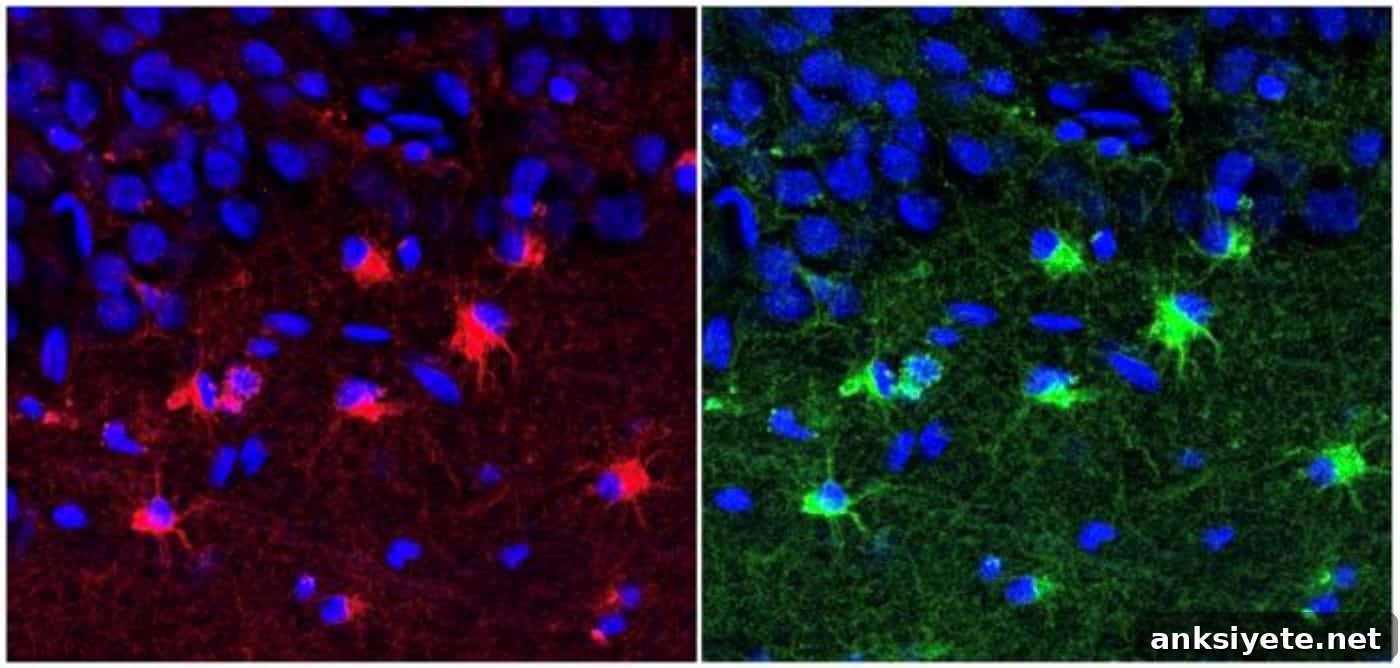
Summary:
USC researchers analyzing living hippocampal tissue removed during epilepsy surgery found that a higher density of abnormal, immature astroglia in the hippocampus is associated with poorer fluid intelligence — the capacity to reason, solve novel problems, and adapt to unfamiliar situations. This direct cellular evidence from living human tissue highlights a non-neuronal driver of complex cognition and points to astroglia as a possible target to slow or treat age-accelerated cognitive decline.
Key Facts:
- Direct human tissue measurements: Researchers quantified immature astroglial cells in hippocampal samples surgically removed from 29 patients with drug-resistant mesial temporal lobe epilepsy, providing rare cellular-resolution data from living human brains.
- Specific cognitive association: A greater prevalence of abnormal immature astroglia correlated specifically with worse performance on fluid intelligence tests, rather than uniform decline across all cognitive domains.
- New glial target for aging: Because fluid intelligence typically declines with normal aging and is affected early in conditions such as frontotemporal dementia, Parkinson’s disease, and multiple sclerosis, promoting astroglial maturation or removing dysfunctional glia could be a novel strategy to preserve reasoning abilities.
Source: Keck School of Medicine of USC
Fluid intelligence — the ability to think logically, identify patterns, and solve unfamiliar problems independent of learned knowledge — declines with age and is among the first cognitive functions to be impaired in several neurodegenerative disorders.
Historically, declines in reasoning and problem solving have been attributed mainly to neuronal loss or synaptic dysfunction. The role of glial support cells in human cognitive aging has been harder to assess directly because most approaches rely on imaging with limited cellular resolution or on post-mortem tissue that lacks recent standardized cognitive testing.
A team led by researchers from the USC Stem Cell laboratory of Michael Bonaguidi, PhD, and the USC Neurorestoration Center investigated this question by combining detailed histology with pre-surgical neuropsychological testing in patients undergoing therapeutic hippocampal resections.
Rare access to living hippocampal tissue
Non-invasive tools such as MRI and EEG cannot resolve distinct glial cell states at a cellular level, and post-mortem specimens often suffer from degradation and the absence of recent cognitive assessments. The USC investigators obtained a unique dataset by examining hippocampal tissue removed from 29 patients with mesial temporal lobe epilepsy (MTLE). Each patient completed comprehensive neuropsychological batteries before surgery that evaluated reasoning, working memory, attention, language, and motor skills. Immediately after resection, researchers performed microscopic analysis to quantify immature astroglia across defined hippocampal regions.
The link between immature astroglia and cognitive decline
Astroglia are star-shaped glial cells that provide metabolic support, maintain the blood-brain barrier, regulate neurotransmitter recycling, and contribute to synaptic homeostasis. In this study, tissue analysis showed that older patients had a higher burden of astroglia arrested in an abnormal, immature state. When these cellular measurements were compared with patients’ pre-surgical cognitive scores, a clear pattern emerged: patients with greater numbers of immature, abnormal astroglia performed worse on tests of fluid intelligence.
“We found that older patients tended to have more abnormal immature astroglia, and that higher numbers of these cells were also associated with poorer performance on tests of fluid intelligence,” said first author Aswathy Ammothumkandy, PhD. These results indicate that astroglia may influence the brain networks that underlie reasoning and flexible problem solving.
Opening a new glial frontier for drug discovery
Because the immature astroglia identified by the team appear functionally arrested, they may fail to support synaptic plasticity and proper network dynamics in the hippocampus, undermining the neural computations required for complex reasoning. Targeting these dysfunctional cells could therefore protect or restore fluid intelligence.
The research group is now screening for molecular markers that distinguish abnormal immature astroglia from healthy mature astrocytes. Defining transcriptional and metabolic signatures may enable the development of small molecules that promote astroglial maturation or selectively eliminate dysfunctional cells. The team also plans to test whether targeted neuromodulation — precise electrical or magnetic stimulation that shifts hippocampal network states — can encourage healthy astroglial physiology and preserve fluid reasoning across the lifespan.
Funding:
This work was supported by the National Institutes of Health (R01AG076956), the Cure Alzheimer’s Fund, The Eli and Edythe Broad Foundation, the USC Neurorestoration Center, the Rudi Schulte Research Institute, the American Epilepsy Society, the California Institute for Regenerative Medicine (EDUC4-12756), and CURE Epilepsy Taking Flight.
Editorial notes:
- The article was edited by staff with expertise in neuroscience reporting.
- The journal paper was reviewed in full by the editorial team.
- Additional context and clarifications were added to support reader understanding.
About this neuroscience research:
- Media contact: Laura LeBlanc
- Source: USC
- Image credit: Aswathy Ammothumkandy / Bonaguidi Lab / USC Stem Cell
- Original research (open access): Frontiers in Cellular Neuroscience (Oct 19, 2026). Title: “An age-related decline in human fluid intelligence corresponds with immature astroglia prevalence.” Authors include Aswathy Ammothumkandy and colleagues.
- DOI: 10.3389/fncel.2026.1926804
Abstract
An age-related decline in human fluid intelligence corresponds with immature astroglia prevalence
Age-related cognitive decline remains a major public health challenge, and strategies focused solely on neurons have not halted its progression. Astroglia are increasingly recognized as important contributors to cognitive function and dysfunction, but their role in human age-associated cognitive decline has been unclear. Patients with mesial temporal lobe epilepsy exhibit progressive, age-amplified cognitive decline, offering an opportunity to examine cellular contributors.
Building on prior work that identified pathological Dcx-positive immature astroglia in epileptic hippocampus, this study combined histological measurements and pre-surgical neuropsychological data to evaluate (1) age-associated cognitive decline in MTLE patients (N = 40), (2) age-related changes in Dcx-positive immature astroglia (N = 29), and (3) the association between immature astroglia and cognitive decline (N = 19).
Results showed a marked decline with age in fluid intelligence, visuospatial reconstruction, and memory. An increase in Dcx-positive immature astroglia correlated positively with age and negatively with nonverbal abstract reasoning and fluid intelligence as measured by Raven’s Progressive Matrices. These observations suggest a link among aging, reduced fluid intelligence, and the prevalence of immature astroglia in epilepsy patients, supporting the idea that immature astroglia are a cellular target worth pursuing to modify age-associated cognitive decline.