
Summary:
Researchers at the University of Oxford have shown that a specialized subset of human microglia can precisely remove toxic alpha-synuclein aggregates from living dopamine-producing neurons without destroying the host cells. Using co-cultures of human induced pluripotent stem cell (iPSC)-derived dopaminergic neurons and microglia, the team demonstrated a targeted “trogocytosis” mechanism—cellular nibbling—driven by GPNMB, regulated by P2RY12 and CD22 signaling, and restrained by an IL-10 autocrine brake. These results identify a neuroprotective microglial phenotype with potential implications for Parkinson’s disease therapies.
Key Facts:
- Selective pruning by trogocytosis: Human microglia remove membrane-enclosed pockets of aggregated alpha-synuclein from intact dopaminergic neurons, preserving neuronal structure and connectivity.
- GPNMB as a functional driver: Glycoprotein non-metastatic melanoma protein B (GPNMB), previously linked to Parkinson’s disease risk in GWAS, binds pathological alpha-synuclein and is required for efficient microglial clearance.
- Balanced immune control: The clearance process is coordinated by P2RY12 sensing, CD22 inhibitory interactions, and an IL-10-mediated feedback loop that limits excessive inflammatory activation.
Source: University of Oxford
Beyond the Double-Edged Sword: A Precision Immune Defense
Microglia are the brain’s resident immune cells and are often described as having both helpful and harmful roles in neurodegeneration. Under normal conditions they survey the brain, prune synapses and clear debris. In chronic disease, prolonged microglial activation can promote inflammation that worsens neuronal injury.
This dichotomy is particularly relevant to Parkinson’s disease, which affects millions worldwide and is marked by the progressive loss of substantia nigra dopaminergic neurons. A major driver of this degeneration is the misfolding and aggregation of the neuronal protein alpha-synuclein into toxic intracellular inclusions.
Until now, it was unclear whether human microglia could remove these intraneuronal aggregates in a way that spares the affected neurons. The University of Oxford team identified a precise mechanism by which a defined microglial population accomplishes just that.
Instead of engulfing and destroying whole neurons, these microglia selectively “nibble” away membrane regions that contain aggregated alpha-synuclein, a process known as trogocytosis. This targeted removal reduces the burden of toxic protein while preserving the neuron’s axons and cell body, suggesting a protective role in early disease stages.
Engineering Human iPSC Co-Cultures to Model Live Interactions
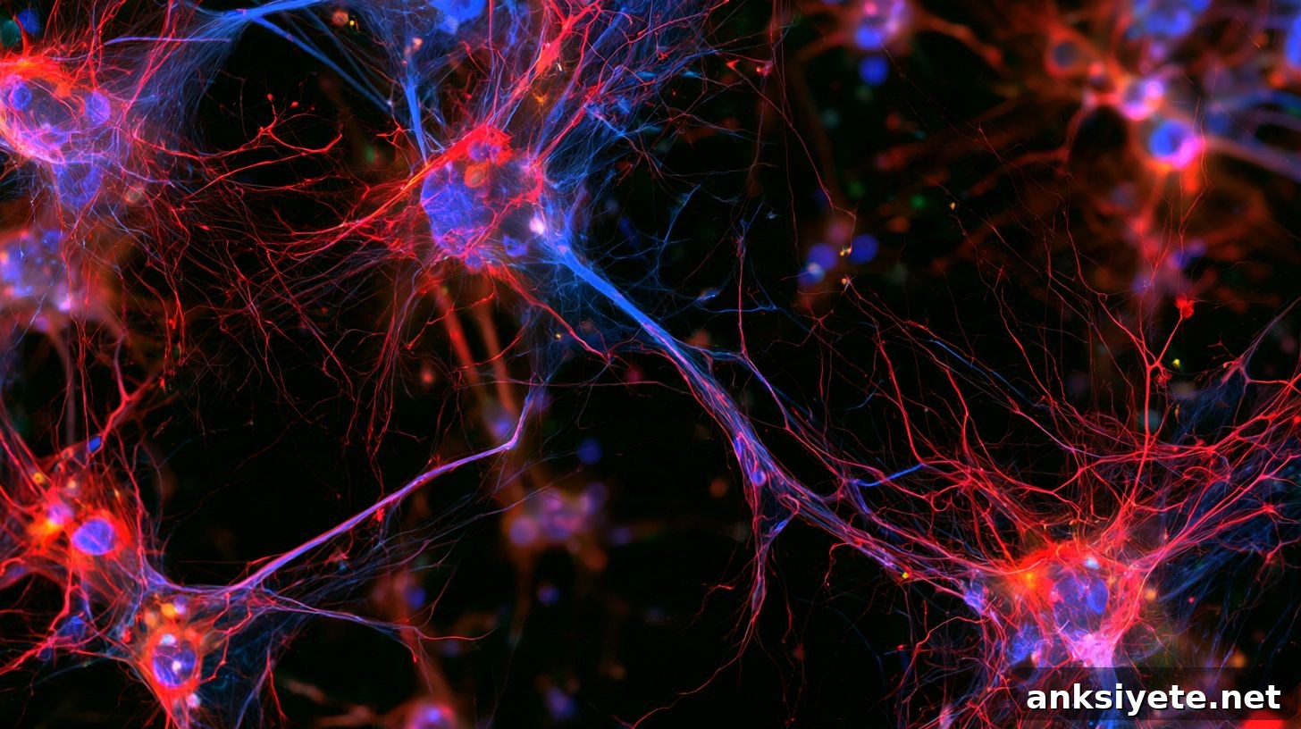
To study human cell interactions directly, investigators created co-cultures from human iPSC lines that generate authentic dopaminergic neurons alongside human microglia. They induced alpha-synuclein pathology using two complementary strategies: increasing alpha-synuclein gene dosage (triplication) and seeding cultures with preformed alpha-synuclein fibrils that template misfolding.
High-resolution live imaging showed that microglia make direct contact with affected neurons and extract membrane-enclosed aggregates rather than performing wholesale phagocytosis of dying cells. Single-cell RNA sequencing revealed that this activity is carried out by a transcriptionally distinct subset of activated microglia—disease-associated microglia (DAM)—that acquire a specific gene-expression program enabling trogocytosis and lysosomal degradation of aggregates.
The microglial response is tightly controlled by multiple signals:
- Surveillance: P2RY12 purinergic receptor sensing helps microglia detect and home to affected regions.
- Inhibitory checkpoints: CD22-mediated interactions limit overactivation and prevent inappropriate neuronal loss.
- Anti-inflammatory brake: An IL-10 autocrine loop reduces secondary inflammatory damage and constrains the phagocytic program.
Solving the GPNMB Genetic Mystery in Parkinson’s
Genome-wide association studies had repeatedly implicated the GPNMB gene locus as a Parkinson’s risk factor, but its role in the brain was unclear. This study links GPNMB mechanistically to microglial clearance of alpha-synuclein: GPNMB expression increases in microglia exposed to aggregate-bearing neurons, it physically associates with phosphorylated alpha-synuclein within microglial compartments, and its levels are higher in substantia nigra microglia from individuals with incidental Lewy body pathology and Parkinson’s disease.
Using CRISPR interference to reduce GPNMB expression specifically in microglia impaired their ability to clear intraneuronal aggregates, establishing GPNMB as an essential effector of the neuroprotective trogocytic response.
As senior author George Tofaris, M.D., Ph.D., emphasizes, these findings refine our understanding of immune involvement in Parkinson’s: microglial activity can be beneficial if properly regulated. Identifying and monitoring microglial subtypes that remove pathological protein while avoiding damaging inflammation may offer new avenues for disease-modifying therapies.
Editorial Notes:
- This article was edited by a Neuroscience News editor.
- The original journal paper was reviewed in full.
- Additional context was added by editorial staff.
About this Neurology Research:
- Media Contact: Christopher McIntyre
- Source: University of Oxford
- Image Credit: Image credited to Neuroscience News
- Original Research (Open Access): Science Translational Medicine (Oct 7, 2026). Title: “Human microglia clear intraneuronal alpha-synuclein aggregates by GPNMB-mediated trogocytosis.” Authors: Hung-Ju Chueh et al.
- DOI: 10.1126/scitranslmed.adz9258
Abstract
Human microglia clear intraneuronal alpha-synuclein aggregates by GPNMB-mediated trogocytosis
Microglia are the brain’s principal immune cells, and their role in Parkinson’s disease is complex: prolonged activation can harm neurons, yet microglia can also carry out protective functions early in pathology. Using human iPSC-derived co-culture models, researchers explored how microglia respond to Ser129-phosphorylated alpha-synuclein aggregates that form within dopaminergic neurons from alpha-synuclein gene triplication or are induced by fibrils.
Microglia cleared intraneuronal aggregates via a contact-dependent mechanism characterized as trogocytosis—selective removal of neuronal subcompartments—rather than whole-cell phagocytosis. This process was independent of TREM2 and phosphatidylserine pathways and was modulated by P2RY12 sensing, CD22 inhibitory signaling, and an IL-10 autocrine brake. Single-cell sequencing identified a disease-associated microglial cluster responsible for this phenotype, and GPNMB emerged as a key lysosomal effector that binds phosphorylated alpha-synuclein and promotes aggregate degradation. Microglia-specific knockdown of GPNMB reduced clearance, while blocking IL-10 signaling enhanced phagocytic activity. Together, these data define a human microglial subtype capable of removing intraneuronal aggregates and point to potential therapeutic targets for Parkinson’s disease.