
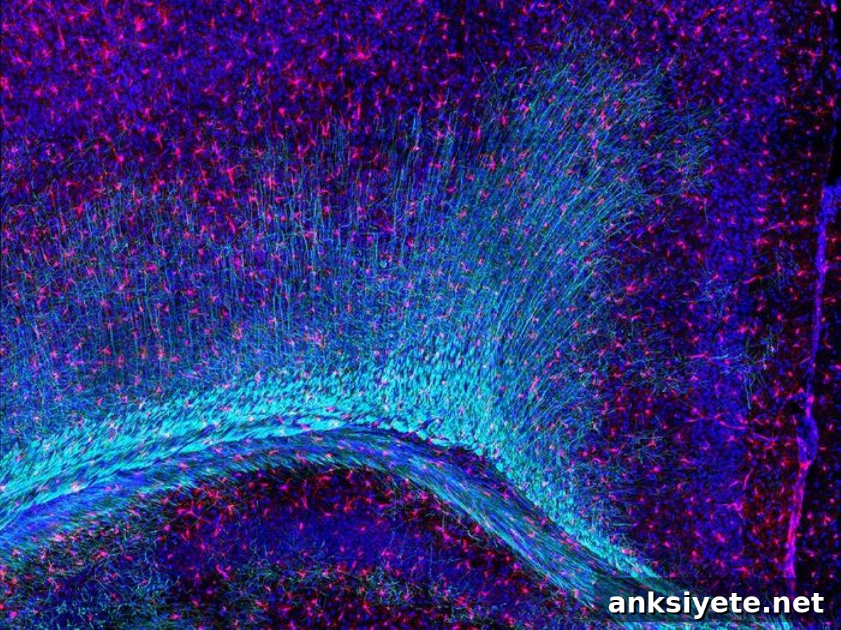
Summary: Researchers have shown that losing normal TDP-43 function specifically in microglia disrupts brain development. Without TDP-43, microglia experience a collapse of the TREM2–DAP12 signaling pathway and can no longer clear routine myelin errors produced during early wiring. This breakdown of neuro-immune housekeeping damages oligodendrocytes, impairs myelin integrity, and produces lasting motor deficits in adult animals.
Key Facts
- Microglia as developmental guardians: Microglia are the brain’s resident immune cells and maintenance crew. During early development they remove dead cells, fight infection, and clear cellular debris to support accurate circuit formation.
- Cross-cell consequences: Deleting TDP-43 in developing microglia causes defects beyond those cells. High-resolution imaging revealed structural alterations in specific brain regions and clear abnormalities in myelin, the insulating sheath that speeds neuronal signaling.
- Oligodendrocyte impact: Oligodendrocytes, the cells that synthesize and repair myelin, are indirectly harmed when microglia fail to maintain a healthy environment. The accumulation of debris and the altered molecular milieu are linked to oligodendrocyte dysfunction.
- TREM2–DAP12 signaling failure: Normal myelination naturally generates small structural errors that microglia recognize and remove via the TREM2–DAP12 axis. Loss of microglial TDP-43 impairs this pathway, leaving microglia unable to detect and clear myelin debris.
- Long-term motor deficits: When microglial housekeeping is compromised during early life, unresolved myelin damage accumulates and disrupts neural transmission. Mice lacking microglial TDP-43 in early development subsequently develop measurable motor and behavioral impairments in adulthood.
Source: University of Lausanne
Rosa Chiara Paolicelli’s research team in the Department of Biomedical Sciences at the University of Lausanne identified a new role for the TDP-43 protein in regulating microglial function. Their work demonstrates how loss of TDP-43 in microglia can contribute to neurodevelopmental disruption and to mechanisms relevant for neurodegenerative disease.
The study centers on microglial biology. Although microglia are best known as the brain’s immune sentinels, they also perform essential maintenance tasks throughout development. Paolicelli’s group used mouse models in which the gene encoding TDP-43 was selectively deleted in microglial cells. Mice that lacked microglial TDP-43 from early life showed structural brain changes and developed motor deficits as adults.
Their findings were published in the 8 July 2026 issue of Nature Neuroscience.
The brain’s guardians and TDP-43
Microglia are small glial cells residing in the brain, spinal cord and retina. Beyond immune surveillance and pathogen defense, they are responsible for clearing dead cells and pruning debris to preserve circuit precision. The Lausanne team focused on TDP-43, a protein implicated in neurodegenerative diseases such as amyotrophic lateral sclerosis (ALS) and some dementias. In disease, TDP-43 relocates from the nucleus to the cytoplasm and can form toxic aggregates, producing loss of normal nuclear functions.
Much of the prior research on TDP-43 has centered on neurons. Paolicelli’s study addresses the less explored question of how TDP-43 loss affects microglia and, by extension, brain development and long-term function.
“We removed TDP-43 specifically from microglia to observe the downstream effects,” explains Rosa Chiara Paolicelli. “The results show that microglial loss of TDP-43 during early development is sufficient to induce lasting motor impairments.”
Multilevel harm revealed by imaging and molecular analysis
Using magnetic resonance imaging alongside confocal and electron microscopy, the researchers identified structural changes in discrete brain regions during early postnatal development and widespread myelin abnormalities. Spatial transcriptomics uncovered a signature of interferon-responsive genes and molecular patterns consistent with oligodendrocyte stress and dysfunction.
Functionally, microglia lacking TDP-43 showed impaired capacity to engulf and degrade myelin debris. At the molecular level, loss of TDP-43 induced cryptic exon inclusion in Tyrobp mRNA, producing truncated DAP12 protein and impairing TREM2 signaling—disabling the pathway microglia use to sense and remove myelin damage.
“These results reveal an unexpected role for TDP-43 in maintaining the TREM2–DAP12 axis and microglial housekeeping,” says Anne-Claire Compagnion, first author of the study. “When that system fails, the secondary effects on oligodendrocytes and myelin compromise neural circuitry and behavior.”
Key Questions Answered:
A: Oligodendrocytes build myelin, but the myelination process naturally produces small structural errors and debris during childhood. Microglia act as the clean-up crew, removing these imperfections so myelin remains functional. If microglia lose TDP-43 and can no longer perform this maintenance, debris accumulates, creating an environment that impairs oligodendrocytes and leads to broader myelin dysfunction.
A: The TREM2–DAP12 axis is a key signaling pathway microglia use to detect and respond to tissue damage and debris. TREM2 senses extracellular cues and signals through DAP12 to trigger engulfment and degradation. The study shows that loss of microglial TDP-43 disrupts this signaling, leaving microglia unable to clear myelin debris effectively.
A: Historically, disorders such as ALS and some dementias have been viewed primarily as neuron-centric. This research expands that view by demonstrating a critical non-neuronal role for TDP-43 in microglia. Disruption of microglial function during development or disease could contribute to structural decline and functional loss, pointing to new therapeutic opportunities aimed at preserving immune-cell housekeeping to protect neural circuits.
Editorial Notes:
- This article was edited by a Neuroscience News editor.
- The journal paper was reviewed in full.
- Additional context was provided by staff.
About this research news
Author: Géraldine Falbriard
Source: Stellate Communications, University of Lausanne communications
Contact: Géraldine Falbriard – Stellate Communications
Image: The image is credited to Paolicelli Lab, Unil
Original Research: Open access.
“Microglial TDP-43 mediates myelin refinement and represses Tyrobp cryptic exon inclusion in mice” by Anne-Claire Compagnion et al., Nature Neuroscience. DOI: 10.1038/s41593-026-02348-3
Abstract
Microglial TDP-43 mediates myelin refinement and represses Tyrobp cryptic exon inclusion in mice
Pathological changes in TDP-43 characterize several neurodegenerative disorders, including amyotrophic lateral sclerosis and frontotemporal dementia, where TDP-43 is mislocalized in neurons and glia. The specific role of TDP-43 in microglia and the consequences of its loss remained unexplored.
Combining magnetic resonance imaging with confocal and electron microscopy, the authors identified early postnatal structural changes and myelin abnormalities in mice lacking TDP-43 in microglia. Spatial transcriptomics revealed an interferon-responsive signature associated with oligodendrocyte dysfunction, and early microglial TDP-43 loss produced motor deficits in adult animals.
Mechanistically, TDP-43 knockout impaired microglial uptake and degradation of myelin and caused cryptic exon inclusion in Tyrobp mRNA, yielding truncated DAP12 protein and defective TREM2 signaling. These findings reveal that TDP-43 regulates the TREM2–DAP12 axis in mice and identify a previously unrecognized mechanism by which TDP-43 controls microglial function.