
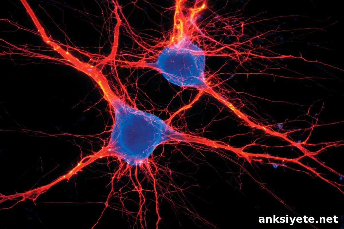
Summary: Scientists have identified a specific subpopulation of epinephrine-producing neurons, called C1 neurons, that act as powerful drivers of fear and persistent anxiety. Under normal conditions, these cells issue a short-lived alarm during intense stress. The new study shows that prolonged, intense activation of this pathway can lock a downstream stress center into a prolonged “on” state, producing severe anxiety that persists for days after the threat has passed.
Key Facts
- Rethinking the brainstem: C1 neurons are located in the rostral ventrolateral medulla (RVLM), a primitive brainstem area historically associated with basic autonomic functions such as breathing and heart rate. Finding neurons there that influence complex emotional states like chronic anxiety challenges prior assumptions about brainstem function.
- The PAG connection: Using a precision-targeting approach developed in the Schwarz lab, researchers isolated C1 neurons and mapped a direct, potent pathway from these cells to the periaqueductal gray (PAG), a central hub that coordinates behavioral responses to stress.
- From short alarm to week-long anxiety: Brief C1 activation supports survival during immediate danger, but sustained, strong stimulation produces a shift in downstream PAG signaling. That alteration results in anxiety behaviors that can last roughly a week after the initial stimulus.
- Broken off-switch model: The team proposes that normally the C1-to-PAG circuit turns off after a stressor ends. A sufficiently intense or prolonged traumatic event can overstimulate C1 neurons, effectively breaking that off-switch and keeping stress signaling active.
- Therapeutic intervention: Chemically silencing C1 neurons immediately after a severe stressor prevented the development of lasting anxiety-like behaviors in the subjects, pointing to a potential targeted treatment strategy.
- No autonomic side effects observed: Importantly, manipulating C1 neurons did not alter immediate behavior or basic autonomic functions in the experiments. This suggests a future therapy that targets these cells could reduce persistent anxiety without causing sedation, memory impairment, or cardiovascular effects common to many current medications.
Source: St. Jude Children’s Research Hospital
Overview
Anxiety disorders affect more than 300 million people worldwide. Many brain regions have been implicated in anxiety, but the way those regions connect and interact has been unclear. Researchers at St. Jude Children’s Research Hospital examined these connections and found that epinephrine-producing C1 neurons in mice modulate fear and anxiety. Their work suggests these cells play a central role in converting acute stress into prolonged anxiety and identifies them as a promising target for new treatments.
The research, published in Neuron, shows that while C1 neuron activity normally rises temporarily during stress, prolonged activation leads to elevated anxiety that can endure for several days. Pharmacological inhibition of C1 neurons reduced anxiety-like responses, especially when performed immediately after intense stress.
Lindsay Schwarz, PhD, Department of Developmental Neurobiology, emphasizes that C1 neurons appear to promote anxiety without directly affecting autonomic functions, raising the possibility of therapies that selectively prevent long-term emotional consequences without disrupting bodily homeostasis.
C1 neurons and the stress response
C1 neurons are embedded within the RVLM, a region known for controlling breathing and cardiovascular processes. Historically, that diversity made it difficult to distinguish the roles of individual neuron types in stress-related behavior. Using an intersectional, precision-targeting system developed in the Schwarz laboratory, the team selectively probed C1 neurons and demonstrated that their activation excites neurons in the PAG, a key regulator of behavioral and physiological stress responses.
Activation of C1 neurons produces immediate anxiety-like responses in mice. Critically, when activation is strong and prolonged, it sustains PAG activity and extends anxiety for up to a week. The investigators propose that an overstimulated C1-to-PAG circuit fails to deactivate after the threat, leaving the brain in a heightened state of alarm for an extended period.
When researchers inhibited C1 neurons during periods of high stress, the mice were protected from developing later, chronic anxiety-like behaviors. Notably, this inhibition had no observable effect on in-the-moment behavior, heart rate, or breathing, supporting the idea that therapeutically targeting these neurons could reduce persistent anxiety without impairing normal responses to danger or bodily function.
Authors and funding
First author: Carlos Fernández-Peña (formerly St. Jude, now University of Nebraska Medical Center). Other contributors: Rachel Pace, Lourds Fernando, Heather Sheppard, Brittany Pittman, and Lindsay A. Schwarz, all affiliated with St. Jude.
Funding: Supported by the Brain & Behavior Research Foundation, the National Institutes of Health (1DP2NS115764), and the American Lebanese Syrian Associated Charities (ALSAC).
Key Questions Answered
A: For decades, research on anxiety emphasized higher-order brain regions such as the amygdala and prefrontal cortex. The lower brainstem, including the medulla, was generally seen as responsible for automatic life-support functions. Discovering that a small group of neurons within this primitive area can influence complex, long-lasting emotional states challenges that long-standing view of brain organization.
A: The proposed mechanism is a failure of the circuit’s off-switch. Normally, C1 neurons fire to help an organism respond to danger and then quiet down. If the initial stress is intense or prolonged, C1 neurons can become overactive and keep driving the downstream PAG pathway. That persistent signaling sustains anxiety even after the environment has returned to safety.
A: In these experiments, inhibiting C1 neurons did not change immediate behavioral responses to stress or basic autonomic parameters like heart rate and respiration. Instead, blocking these cells prevented acute stress from consolidating into long-term anxiety. This suggests a therapeutic strategy that reduces persistent trauma without blunting natural fear or undermining physiological health.
Editorial Notes:
- This article was edited by a Neuroscience News editor.
- The journal paper was reviewed in full.
- Additional context was provided by editorial staff.
About this anxiety and neuroscience research news
Author: Chelsea Bryant
Source: St. Jude Children’s Research Hospital
Contact: Chelsea Bryant – St. Jude Children’s Research Hospital
Image: The image is credited to Neuroscience News
Original Research: Open access. “Autonomic C1 neurons promote anxiety via activation of vlPAG” by Brittany G. Pittman, Carlos Fernández-Peña, Heather Sheppard, Lindsay A. Schwarz, Lourds M. Fernando, Rachel L. Pace. Neuron
DOI: 10.1016/j.neuron.2026.06.012
Abstract
Autonomic C1 neurons promote anxiety via activation of vlPAG
Anxiety is an emotional state driven by anticipation of threat. While acute anxiety can enhance survival by increasing vigilance and readiness, excessive or persistent anxiety impairs health and quality of life. Despite extensive study, the mechanisms underlying anxiety disorders remain incompletely understood, limiting targeted therapeutic advances.
Using intersectional tools, the authors show that a small population of adrenergic, autonomic brainstem neurons—C1 cells—promote anxiety-related behaviors. Calcium imaging indicates these neurons activate during stressful events, and their inhibition reduces fear responses and prevents the escalation of anxiety. These results identify C1 neurons as a key brain–body hub for the promotion of anxiety-related behaviors and suggest that targeted inhibition of this circuit may offer therapeutic benefit.