
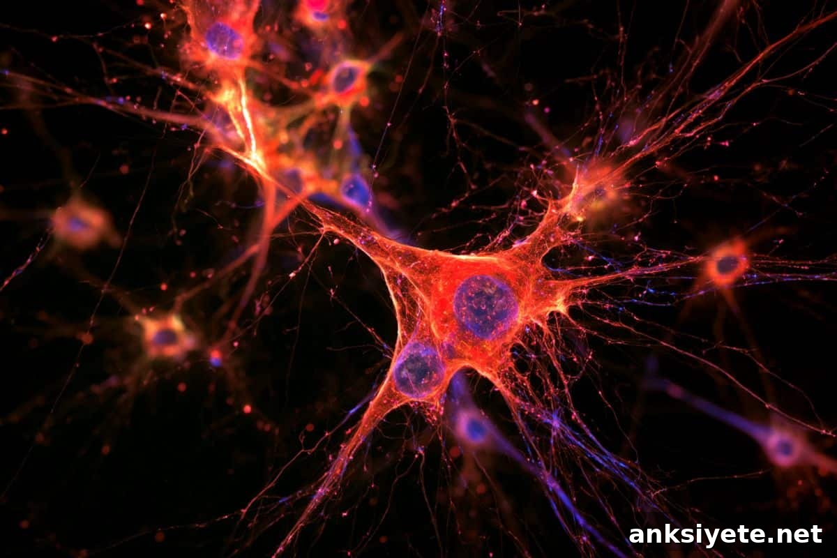
Summary: New research shows that human microglial cells develop far more slowly than those of other species, a delay linked to human-specific duplications of the SRGAP2 gene.
Scientists report that human microglia require roughly four to eight years to reach full maturity, compared with about three weeks in mice. This extended developmental timing—neoteny—aligns microglial maturation with the prolonged development of human neurons, suggesting coordinated timing between immune cells and neural circuits during childhood.
Because microglia actively prune synapses and adjust circuit responsiveness during development, their synchronized neoteny with neurons may be an important evolutionary mechanism that supports complex human cognition and also sheds light on the origins of neurodevelopmental and neurodegenerative disorders.
Key Facts
- Microglial neoteny: Human microglia display an extreme developmental delay, taking four to eight years to mature fully; by contrast, mouse microglia mature in roughly three weeks.
- SRGAP2 duplication in microglia: Human-specific duplicates of the SRGAP2 gene are found at much higher levels in microglia—nearly ten times more abundant than in neurons.
- Synchronized developmental timing: Expression of duplicated SRGAP2 copies helps synchronize microglial maturation with the slow-developing human neurons, enabling coordinated synapse formation and pruning throughout childhood.
- Role in synaptic refinement: During brain development, microglia constitute 5–10 percent of brain cells and actively select which synapses to keep or eliminate, while modulating the responsiveness of neural circuits.
- Relevance to disease: Given microglia’s roles in neurodevelopmental conditions such as autism spectrum disorder and in neurodegenerative diseases like Alzheimer’s, human-specific microglial timing offers an important baseline for understanding disease processes.
Source: Zuckerman Institute
Microglia are the brain’s primary immune cells. They defend against pathogens, clear damaged neurons, and—importantly during development—sculpt neural circuits. Researchers at Columbia’s Zuckerman Institute have now found that human microglia mature unusually slowly compared with those in other species, mirroring the prolonged maturation of human neurons.
“This prolonged development may allow human microglia to influence brain wiring in ways that support our advanced cognitive abilities,” said Carlos Diaz-Salazar, PhD, lead author of the new study published in Neuron, conducted while he worked in the lab of Franck Polleux, PhD.
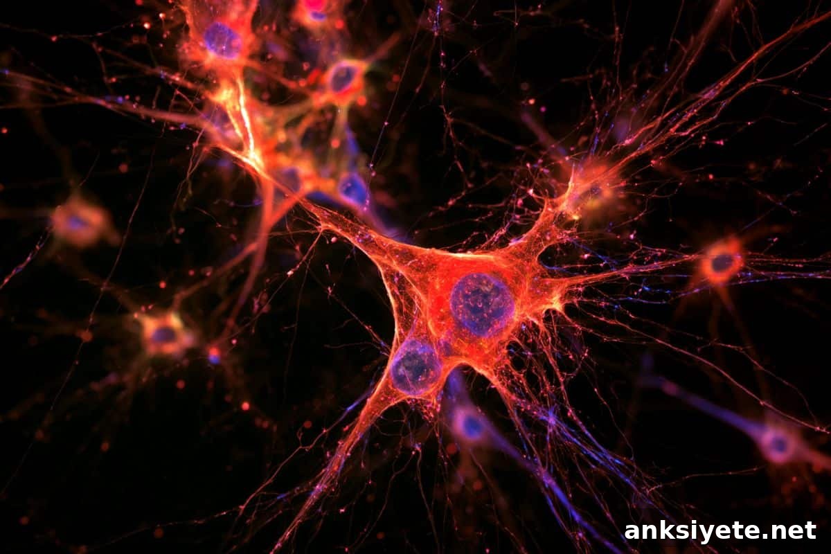
The Polleux lab has focused for more than 15 years on SRGAP2, one of several genes duplicated only in humans. Earlier work from the lab showed that human-specific SRGAP2 duplicates increase the number of synaptic connections neurons form and delay synapse maturation—features that contribute to human neurons’ distinctive strength, density, and capacity to process and store information.
In the current study, Diaz-Salazar and colleagues first found that human-specific SRGAP2 duplicates are nearly ten times more abundant in microglia than in neurons. That observation raised a central question: why is SRGAP2 so active in microglia?
Microglia make up about 5–10 percent of brain cells. Beyond immune defense and debris clearance, a growing body of research shows microglia play an essential role in brain development: they help determine which synapses persist and which are eliminated, and they fine-tune the responsiveness of circuits.
Using a combination of mouse models and human cell-based experiments, the researchers demonstrated that human-specific SRGAP2 duplicates extend microglial maturation to four to eight years, whereas mouse microglia mature in approximately three weeks. The team concludes that SRGAP2 influences the developmental tempo of both neurons and microglia so these cell types mature in concert.
“SRGAP2 helps control the pace of neuronal development, and natural selection appears to have co-opted it to slow microglial maturation as well,” said Diaz-Salazar, who is now at the Hospital del Mar Medical Research Institute in Barcelona. “That synchronization likely ensures microglia are matched to the prolonged period during which human neurons form, refine, and stabilize connections.”
Human brain neoteny—the unusually long window of brain development—is widely considered a foundation for humans’ enhanced cognitive abilities. The team now aims to unravel the cellular and molecular mechanisms by which SRGAP2 duplicates promote neoteny in neurons, microglia, and other brain cell types.
“By identifying the elements that shape the human brain’s prolonged development, we can better understand what makes our species unique from an evolutionary perspective,” said Dr. Polleux. “Because microglia are increasingly implicated in both developmental and degenerative brain disorders, clarifying how human microglia differ from those of other species also advances our understanding of disease mechanisms.”
Key Questions Answered:
A: Microglia are the brain’s resident immune cells, comprising about 5–10 percent of brain cells. In development they not only defend the brain and clear debris but also actively shape neural circuits by pruning synapses and adjusting circuit responsiveness.
A: Neoteny describes a prolonged developmental timeline. Human-specific duplicates of SRGAP2 slow maturation of both neurons and microglia, extending key phases of brain development from weeks (in other species) to years in humans.
A: Because microglia control synaptic pruning and circuit refinement while neurons take years to establish stable, dense connections, synchronized maturation ensures precise sculpting of the complex networks needed for human cognitive function.
Editorial Notes:
- This article was edited by a Neuroscience News editor.
- The journal paper was reviewed in full.
- Additional context was provided by the editorial staff.
About this neurotech and memory research news
Author: Charles Choi (Zuckerman Institute)
Source: Zuckerman Institute
Contact: Charles Choi – Zuckerman Institute
Image: Image credit: Neuroscience News
Original Research: The findings are reported in the journal Neuron.