
Summary: Loss of the sense of smell (anosmia or hyposmia) is a frequent early sign of Parkinson’s disease and can appear years before motor symptoms. New anatomical findings help explain why smell deteriorates in people with Parkinson’s.
Source: University of Auckland
Researchers identify an anatomical deficit in the olfactory bulb linked to smell loss in Parkinson’s disease
Researchers at the University of Auckland have identified a structural change in the human olfactory bulb that correlates with the smell loss commonly observed in Parkinson’s disease. The study, published in the journal Brain, reports a marked reduction in the glomerular component of the olfactory bulb in people who had Parkinson’s, providing an anatomical explanation for early olfactory dysfunction in this neurodegenerative condition.
The study was led by Associate Professor Maurice Curtis, Dr Victor Dieriks, Dr Sheryl Tan and Sir Richard Faull from the Centre for Brain Research at the University’s Faculty of Medical and Health Sciences. The team collaborated with Dr Peter Mombaerts and Dr Bolek Zapiec from the Max Planck Research Unit for Neurogenetics in Frankfurt, Germany. Funding came from the Neuro Research Charitable Trust in New Zealand and the Max Planck Society in Germany.
Parkinson’s disease is a progressive neurological disorder characterized by tremor, rigidity, and slowed movement. While much attention focuses on motor symptoms, olfactory dysfunction is a highly prevalent and early non-motor symptom. According to clinical data, a diminished or lost sense of smell affects the majority of early-stage Parkinson’s patients and often appears years before noticeable movement problems.
The olfactory bulb is the first central brain structure that processes scent information received from olfactory sensory neurons in the nasal cavity. Sensory neuron axons converge into spherical structures called glomeruli. Each glomerulus acts as a functional unit where odor signals are initially processed and then relayed to other brain regions involved in smell perception, including the olfactory cortex.
Comparing postmortem olfactory bulbs from people with and without Parkinson’s disease, the researchers discovered a dramatic decrease in the volume occupied by glomeruli in Parkinson’s cases. Quantitative analyses showed that the global glomerular volume in Parkinson’s olfactory bulbs was reduced by more than half compared with normal controls. In addition to reduced overall glomerular volume, the spatial distribution of glomeruli was altered: while normal olfactory bulbs contained about 70 percent of their glomerular component in the ventral (lower) half, Parkinson’s cases showed only about 44 percent in that region.
These findings support the hypothesis that environmental agents—such as bacteria, viruses or toxins—may enter the brain through the nose, first affecting the olfactory bulb. A ventral-predominant glomerular deficit is consistent with a model in which pathogenic processes begin in the nasal cavity and spread into the brain through olfactory pathways, triggering or contributing to the neurodegenerative cascade of Parkinson’s disease.
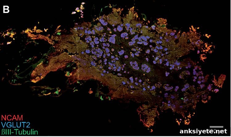
The international team assembled an extensive collection of olfactory bulbs suitable for detailed, quantitative study. Researchers processed entire olfactory bulbs postmortem, sectioning them into thousands of horizontal slices only ten micrometers thick. The sections were stained with fluorescent antibodies in New Zealand, scanned in Frankfurt with a fluorescence slide scanner, and reconstructed in three dimensions to enable whole-olfactory-bulb quantitative analysis.
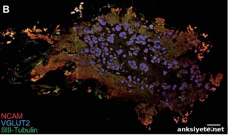
Because individual human glomeruli can be difficult to count precisely, the team developed a robust, quantitative metric called the global glomerular voxel volume. This measure represents the total volume of all voxels identified as glomerular by immunohistochemical labeling in the 3D reconstructions. Using this objective parameter, the researchers demonstrated a substantial loss of glomerular volume in Parkinson’s disease. Whether this reduction reflects fewer glomeruli, smaller glomeruli, or both remains to be clarified by further study.
The Neurological Foundation of New Zealand Douglas Human Brain Bank at the University of Auckland coordinated ethical tissue collection, working closely with families to obtain well-preserved postmortem samples from people with Parkinson’s disease and matched controls. High-quality human tissue enabled the rigorous whole-bulb reconstruction and volumetric analyses reported in the study.
These results will guide further investigations into the mechanisms underlying glomerular deterioration in Parkinson’s disease. Ongoing follow-up studies aim to determine what triggers glomerular loss, how glomerular changes relate to α-synuclein pathology, and what additional structural or cellular alterations occur in the olfactory bulb as the disease progresses. Ultimately, a clearer understanding of early olfactory changes may improve early detection strategies and shed light on disease origins.
Publication: The study appears in the journal Brain and is titled “A ventral glomerular deficit in Parkinson’s disease revealed by whole olfactory bulb reconstruction.” The authors include Bolek Zapiec, Birger V. Dieriks, Sheryl Tan, Richard L. M. Faull, Peter Mombaerts, and Maurice A. Curtis. DOI: 10.1093/brain/awx208.
Funding and support: Work in New Zealand was supported by the Neuro Research Charitable Trust; work in Germany was supported by the Max Planck Society. Tissue was provided through the Douglas Human Brain Bank, Neurological Foundation of New Zealand.
Abstract (summary)
Olfactory dysfunction is a common and early symptom of Parkinson’s disease, yet its underlying causes are not well understood. The study presents a quantitative whole-olfactory-bulb analysis comparing horizontal 10 µm sections from six normal and five Parkinson’s disease cases, stained for markers of glomeruli. Three-dimensional reconstructions enabled measurement of the total glomerular voxel volume and mapping of glomerular distribution along the dorsal–ventral axis. The global glomerular voxel volume in Parkinson’s disease cases was approximately half that of normal cases, and the typical ventral predominance of glomeruli in normal olfactory bulbs was disrupted in Parkinson’s cases. The glomerular deficit, predominantly ventral, aligns with hypotheses that environmental agents entering via the nose may initiate pathogenic processes in Parkinson’s disease. Increased serine 129-phosphorylated α-synuclein load correlated with lower global glomerular volume. The quantitative approach provides a reproducible framework to study normal human olfactory anatomy and its alterations in Parkinson’s disease.