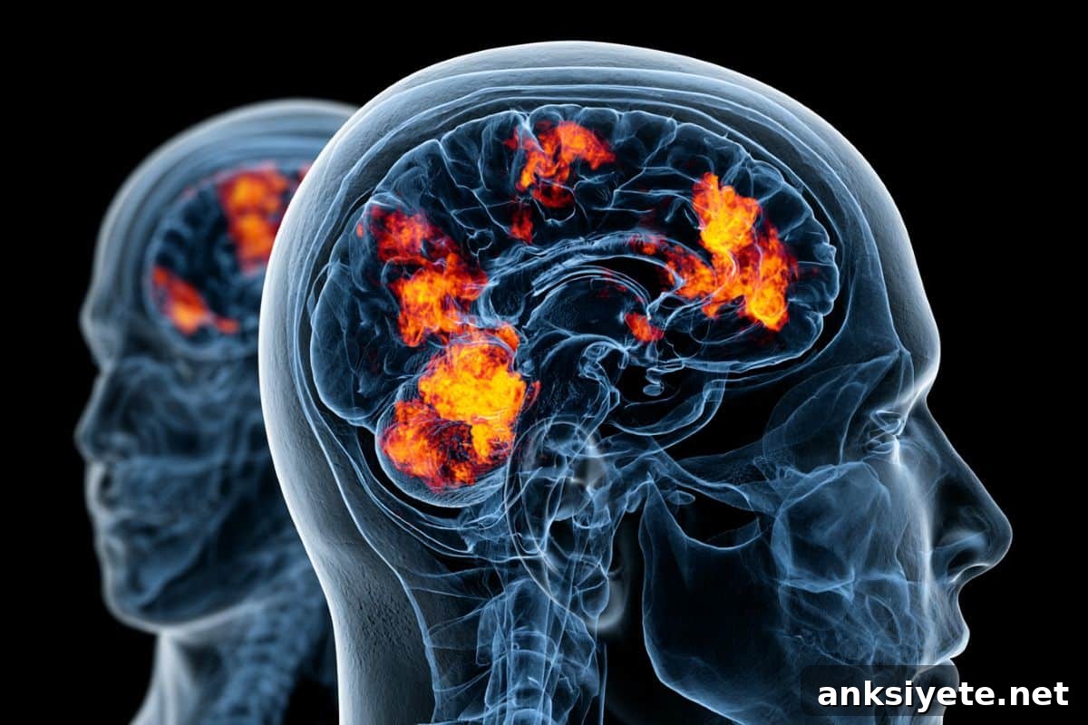
Summary: A randomized clinical trial indicates that using individualized brain imaging to guide transcranial magnetic stimulation (TMS) significantly improves outcomes for people with treatment-resistant depression. Investigators found that selecting stimulation targets from each person’s resting-state functional magnetic resonance imaging (fMRI) connectivity map produced larger antidepressant effects than standard scalp-based targeting.
The study used accelerated TMS (aTMS), a high-dose protocol that delivers multiple daily sessions and compresses a typical multi-week course into one week. By analyzing resting-state functional connectivity to identify how brain regions synchronize at rest, researchers located personalized neural circuit targets for stimulation.
One month after treatment, participants whose aTMS was directed by connectivity-based, individualized fMRI targets experienced substantially greater symptom reductions and higher response rates than those whose targets were determined by conventional scalp measurements. These findings support a more precise, scalable approach to psychiatric neuromodulation.
Key Facts
- Personalized targeting improves outcomes: Choosing aTMS targets using each patient’s fMRI-derived connectivity map provided a measurable advantage over scalp-based methods that do not account for individual brain-circuit differences.
- Condensed treatment schedule: Accelerated TMS (aTMS) delivers multiple stimulation sessions each day, reducing a typical multi-week course to a single intensive week.
- Higher remission and response rates: At one month post-treatment, the connectivity-targeted group achieved an 80% response rate versus 60% in the scalp-targeted group.
- Blinded, objective assessment: Symptom change was assessed with the clinician-rated Montgomery-Åsberg Depression Rating Scale (MADRS), and both participants and raters were blinded to targeting assignment.
- Prospective validation: Rather than relying on retrospective analyses, this randomized trial provides prospective evidence that functional neuroimaging can be applied to improve clinical outcomes in aTMS treatment.
Source: Mass General
Mass General Brigham study indicates functional brain imaging can guide accelerated transcranial magnetic stimulation for depression.
A randomized clinical trial led by investigators at Mass General Brigham’s Neuroscience Institute and Department of Psychiatry evaluated whether individualized, connectivity-based targeting with fMRI enhances the antidepressant benefits of accelerated transcranial magnetic stimulation (aTMS) in adults with treatment-resistant major depressive disorder. Results appear in JAMA Psychiatry.
TMS is a noninvasive neuromodulation method that uses magnetic pulses to alter brain activity. FDA clearance for repetitive TMS to treat major depressive disorder in adults dates to 2008, and TMS is commonly used when standard treatments have not achieved remission.
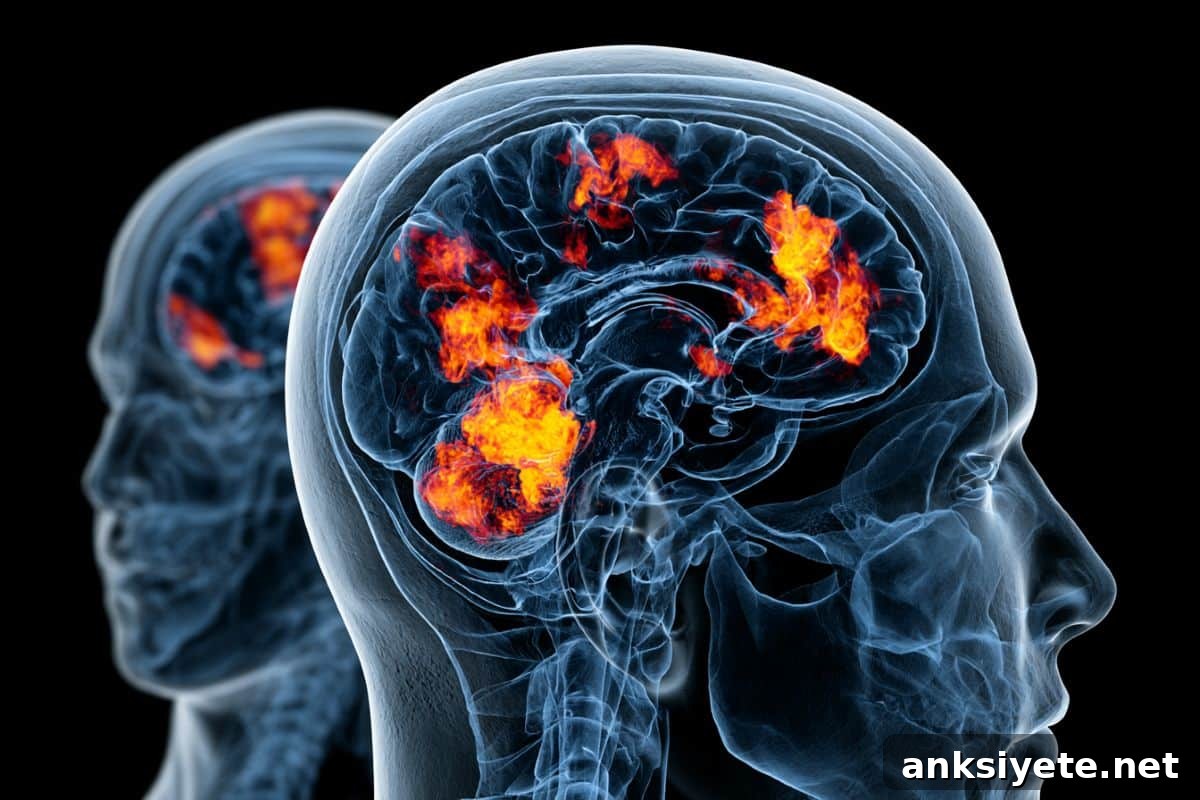
Typically, TMS targets are chosen using scalp-based landmarks. These conventional methods are practical and widely used but do not capture individual differences in the functional circuits implicated in depression. The trial tested whether targeting determined by each participant’s resting-state fMRI connectivity would produce stronger antidepressant responses than the standard scalp-based approach.
The imaging-guided strategy identified the left dorsolateral prefrontal cortex site that showed the strongest correlation with a convergent depression circuit—a network of regions linked to depressive symptoms, including negative connectivity with the subgenual cingulate. The comparison group received targets located using the Beam F3 scalp-based method. All participants received the same aTMS dosing schedule and were blinded to assignment.
“Neuroimaging has greatly expanded our understanding of brain circuits, but demonstrating a direct benefit to patient care has been difficult,” said corresponding author Joseph Taylor, MD, PhD, the Jonathan F. Borus, MD, Endowed Chair in Psychiatry at Mass General Brigham and Assistant Professor of Psychiatry at Harvard Medical School. “Because imaging introduces cost and complexity, our trial measured how much benefit connectivity-based targeting could add beyond conventional approaches.”
Forty adults aged 22 to 80 with major depressive disorder and moderate-to-severe treatment resistance were randomized after a pre-treatment multiecho resting-state fMRI. Participants, TMS technicians, and clinical raters were blinded to targeting method. The primary outcome was MADRS score at one month post-treatment, with longer-term follow-up scheduled every three months for a year.
One month after therapy, the connectivity-targeted group showed significantly larger reductions in MADRS scores than the scalp-targeted group. Median MADRS reduction was 24 points (IQR 19–28) for the connectivity group versus 18 points (IQR 10–23) for the scalp group (P = .02). The estimated effect size was large (analogous to Cohen’s d = 0.8), and individualized targets were reliable within individuals while differing significantly between participants. Clinically, 80% of those receiving connectivity-based targeting met response criteria versus 60% in the scalp-targeted group.
These outcomes build on prior circuit-targeting research from the team and provide prospective evidence that fMRI-guided targeting can enhance the effects of high-dose aTMS. As aTMS becomes more accessible, these findings can inform decisions about incorporating neuroimaging into clinical practice and planning larger confirmatory trials.
The investigators acknowledge limitations, including the modest sample size and single-site design. They plan larger, multisite studies to confirm efficacy across more diverse patient groups and to assess the durability of benefits.
Authorship: In addition to Joseph J. Taylor, authors from Mass General Brigham include Marina R. Kare, Dania Haj-Darwish, Emma Jones, Lauren Sanderson, Sanaz Khosravani, Jessica Leach, Leanna Bomer, Natalie Hall, Nicole Chiulli, Christopher Lin, William Drew, Stephan T. Palm, Anjali Chandra, Summer B. Frandsen, Anastasia Bekou, Tracy Barbour, Sheena R. Baratono, Irene Gonsalvez, Stanley Lyndon, Fredric L.W.V.J. Schaper, David Silbersweig, Shan H. Siddiqi, and Michael D. Fox. Additional contributors include Elizabeth Steuber and Wei Wang.
Disclosures: Taylor reported grants from the Brain and Behavior Research Foundation during the study. Taylor, Fox, and Siddiqi are coinventors on a provisional patent application related to methods described in this trial. Siddiqi and Fox report consultancy fees. Full disclosures appear in the published paper.
Funding: The study was funded by the Brain and Behavior Research Foundation (grant 31081). Additional support for Taylor included awards from Harvard Medical School (Dupont Warren Fellowship Award, Livingston Award), the Sidney R. Baer, Jr. Foundation, the Baszucki Brain Research Fund, Mass General Brigham’s Women’s Brain Initiative and Accelerator Award, and NIH grants (K23MH129829, R01MH113929).
Key Questions Answered:
A: Traditional TMS administers daily sessions spread over several weeks. Accelerated TMS (aTMS) delivers multiple sessions each day to complete an intensive treatment course within a single week. Because aTMS concentrates high doses of stimulation in a short time, fMRI-based targeting helps ensure that this intense neuromodulation is precisely aligned with an individual’s resting-state network organization rather than an approximated scalp location.
A: The trial used the Montgomery-Åsberg Depression Rating Scale (MADRS) to measure symptom change at one month. Participants mapped with individualized connectivity targets experienced significantly larger MADRS score reductions and higher response rates (80% vs 60%) compared with those receiving scalp-based targeting.
A: Main limitations include the small sample size (40 participants) and single-site design. The research team plans larger, multi-center trials to validate efficacy across diverse populations and to examine longer-term outcomes after connectivity-guided aTMS.
Editorial Notes:
- This article was edited by a Neuroscience News editor.
- The journal paper was reviewed in full.
- Additional editorial context was added by staff.
About this TMS and depression research news
Author: Cassandra Falone
Source: Mass General
Contact: Cassandra Falone – Mass General
Image: Image credit: Neuroscience News
Original Research: Open access. “Connectivity- versus scalp-based targeting of accelerated TMS for depression: A randomized trial” by Joseph J. Taylor et al., JAMA Psychiatry. DOI: 10.1001/jamapsychiatry.2026.1100
Abstract
Connectivity- versus scalp-based targeting of accelerated TMS for depression: A randomized trial
Importance
Although neuroimaging is widely used in psychiatric research, evidence that it improves clinical outcomes has been limited. Functional-connectivity imaging has been applied to personalize accelerated TMS (aTMS) and yield rapid antidepressant effects, but the added value of connectivity-based targeting required prospective testing.
Objective
To estimate the effect size of connectivity-guided versus scalp-guided targeting of aTMS for treatment-resistant depression.
Design, Setting, and Participants
This randomized clinical trial compared connectivity- and scalp-based aTMS targeting between July 2023 and March 2025. The primary outcome was assessed at one month posttreatment with follow-up every three months for one year. Participants, TMS technicians, and study clinicians were blinded. The study enrolled 40 adults (age 22–80) with major depressive disorder and moderate-to-severe treatment resistance at the Center for Brain Circuit Therapeutics, Mass General Brigham and Harvard Medical School.
Key exclusions included contraindications to TMS or MRI, primary psychiatric diagnoses other than major depressive disorder or anxiety disorders, recent rapid-acting antidepressant treatments, and significant neurological or substance use disorders.
Interventions
All participants underwent a 41-minute multiecho resting-state functional connectivity scan before aTMS. Half received treatment guided by individualized connectivity targets defined as the left dorsolateral prefrontal cortex site most correlated with a published convergent depression circuit. The comparison used the Beam F3 scalp-based targeting method.
Main Outcomes and Measures
Primary outcome: MADRS score at one month after treatment, adjusted for baseline.
Results
Forty participants (22 female, 55%; mean age 45.7 years) were randomized and treated. Median MADRS reduction was 24 points (IQR 19–28) in the connectivity group versus 18 points (IQR 10–23) in the scalp group (P = .02). The connectivity-based approach yielded an effect-size analogue of 0.8 (95% CI, 0.26–1.54) and a number needed to scan of five. Individualized targets were reproducible within individuals and distinct across participants (split-half distance 4.47 mm vs between-subject distance 12.97 mm; P < .001).
Conclusions and Relevance
Individualized, connectivity-guided targeting of a convergent depression circuit enhanced the antidepressant effects of high-dose aTMS in this randomized trial. These results support cost-effectiveness analyses and help to design a larger confirmatory efficacy trial.
Trial Registration
ClinicalTrials.gov Identifier: NCT05680727