
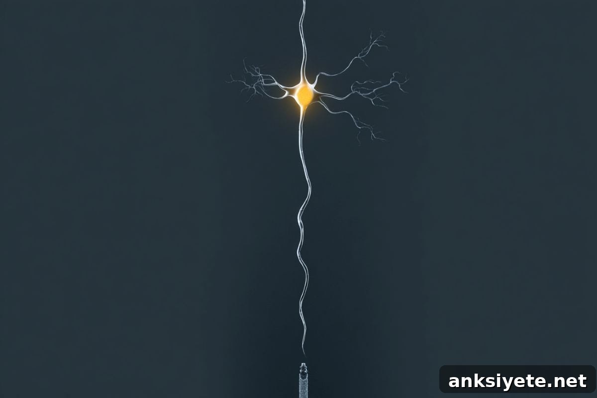
Summary: A major neurotechnology advance now allows scientists to record and manipulate individual neurons simultaneously deep inside the living brain. The device, called Neuropixels Opto, combines large-scale electrophysiology with targeted optogenetic control on a single silicon shank thinner than a human hair.
Validated in mouse models, this next-generation probe delivers high-resolution electrical recordings while enabling precise light-based activation or silencing of specific cells. Early results have already challenged long-standing assumptions about cortical circuit architecture and provide a powerful platform to study the neural circuit disruptions that underlie Alzheimer’s disease, schizophrenia, Parkinson’s disease and other brain disorders.
Key Facts
- The unified neural interface: Neuropixels Opto merges recording and manipulation into one device. Where researchers previously had to choose between listening to electrical signals and delivering optogenetic control, this probe performs both tasks in the same experiment without degrading the electrical signal quality.
- Micro-hair silicon design: The probe integrates roughly 960–1,000 recording sites together with arrays of microscopic light emitters on a single ultra-thin silicon shank. Its narrow form factor lets researchers monitor neural activity across depth while delivering targeted blue and red light stimulation to addressable sites.
- Large-scale collaborative effort: The development is part of a funded, multi-institution project involving teams at UCL, the Allen Institute and international partners. The effort brings together engineering, optics and neuroscience expertise to push high-density neural interfacing forward.
- New view of cortical independence: Using Neuropixels Opto in cortex, researchers observed that neurons can display highly localized activity—operating independently of nearby cells—contradicting expectations that local stimulation would necessarily produce widespread, cascading activation.
- Isolating causal relationships: By recording and manipulating identified cell types in real time, the probe enables experiments that go beyond correlation and reveal how specific neurons causally influence perception, learning and decision-making.
- Relevance for brain disorders: Many neurological and psychiatric conditions reflect circuit-level dysfunctions. This high-resolution, open toolset can help map altered network dynamics in disease models and guide the design of more precisely targeted interventions.
Source: UCL
Overview
Neuropixels Opto integrates high-density extracellular electrophysiology with embedded photonic circuits for optogenetic control. By packing dense arrays of electrical recording sites alongside microscopic light emitters on a hair-thin silicon shank, the device permits simultaneous measurement and manipulation of hundreds of neurons across brain regions, including deep structures.
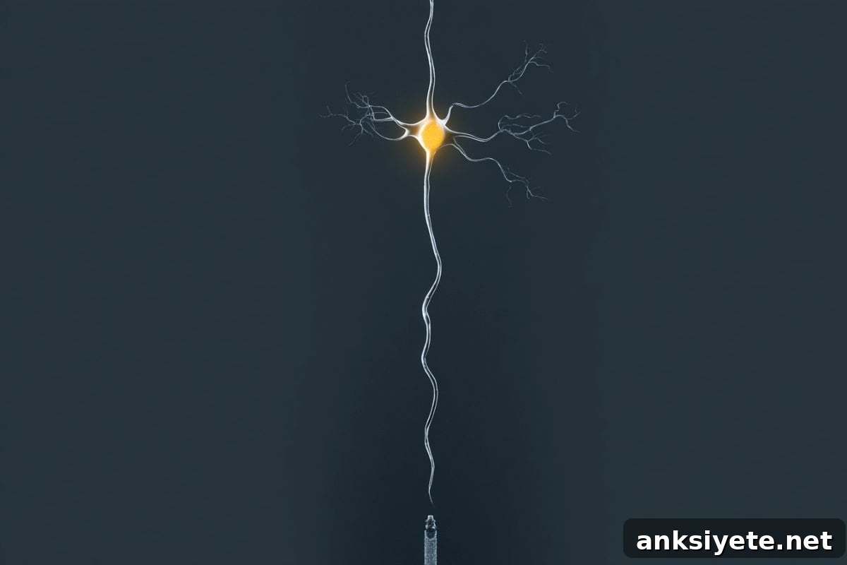
Described in Nature Methods, the system records spikes from distributed neural populations while delivering spatially addressable optogenetic stimuli. In mouse cortex and deeper regions such as the striatum, the probe produced high-quality electrical recordings concurrent with selective activation or silencing of cell types, enabling optotagging and cell-type identification in the same preparation.
Professor Matteo Carandini (UCL Institute of Ophthalmology) explains that understanding how electrical signals across billions of neurons generate behaviour and cognition requires tools that both observe and perturb activity. Neuropixels Opto overcomes technical barriers that previously prevented combining light delivery and sensitive recordings at depth, opening new experimental possibilities for causal circuit mapping.
Probe design and capabilities
The probe’s shank is approximately 70 micrometres wide and can host nearly a thousand electrical sites plus arrays of light emitters arranged to deliver blue and red light at addressable locations. This architecture enables simultaneous multi-site stimulation and recording spanning cortical depths and subcortical targets, offering both single-cell resolution and large-scale network coverage.
Researchers can activate or silence genetically defined cell types while monitoring the real-time response of neighboring neurons along the shank. This combination allows mapping of how specific cells influence local and downstream circuit dynamics, making it possible to test mechanistic hypotheses about information flow, computation and dysfunction in neural networks.
Insights into cortical circuits
Applying Neuropixels Opto in mouse cortex revealed a surprising degree of localization: activating small cell groups did not always trigger broad, synchronous waves as previously assumed. Instead, many neurons could be modulated independently, suggesting finer-grained functional organization and modular processing within cortical circuits.
Key Questions Answered
A: Delivering light deep into tissue creates electrical noise that interfered with sensitive electrodes. Metal electrodes and light sources were difficult to integrate without corrupting recordings, which made simultaneous read-and-write experiments technically infeasible until these engineering challenges were solved.
A: Through advanced microfabrication that integrates electronic and photonic circuits on a sub-100-micrometre silicon platform. Recording nodes and microscopic light emitters are interwoven along the shaft to provide both dense sensing and spatially addressable stimulation.
A: The team observed that many cortical neurons can be modulated in a highly localized manner, operating independently of immediately neighboring cells. This challenges long-held assumptions about inevitable, widespread propagation following local stimulation.
Implications for brain disease research
Because many neurological and psychiatric disorders involve disrupted communication between neurons, a tool that both identifies and manipulates defined cell types while recording network responses is especially valuable. Neuropixels Opto can help delineate how specific circuit elements malfunction in disease models, offering a clearer roadmap for targeted therapeutics and neuromodulation strategies.
Collaborations and broader impact
The project unites laboratories in the UK, US and Europe with engineering partners to advance scalable, high-resolution neural recording technologies. The goal is to make these capabilities more widely accessible to the neuroscience community so researchers worldwide can study circuits at the scale and precision needed to accelerate discovery.
About this research
Author: Henry Killworth
Source: UCL
Contact: Henry Killworth – UCL
Image: Image credit: Neuroscience News
Original Research: Closed access. “Neuropixels Opto: combining high-resolution electrophysiology and optogenetics” by Anna A. Lakunina et al., published in Nature Methods. DOI: 10.1038/s41592-026-03076-z
Abstract
Neuropixels Opto: combining high-resolution electrophysiology and optogenetics
High-resolution extracellular electrophysiology is the standard for recording spikes from distributed neural populations and becomes especially powerful when paired with optogenetics to manipulate specific cell types with millisecond precision. The Neuropixels Opto prototypes integrate electronic and photonic circuits, packing approximately 960 electrical recording sites and two sets of 14 light emitters onto a 70-µm-wide shank. These devices enable spatially addressable optogenetic stimulation using blue and red light while delivering high-quality recordings across cortical and subcortical targets. In mice, the probes provided spatially selective activation and silencing at distinct depths, supported optotagging in deep structures, and represent a promising tool for recording, identifying and manipulating neuronal populations in the same experiment.
Overall, Neuropixels Opto offers a practical path to combine large-scale, high-resolution recording with targeted light-based control—advancing causal circuit analysis and providing a versatile platform for basic and translational neuroscience research.