
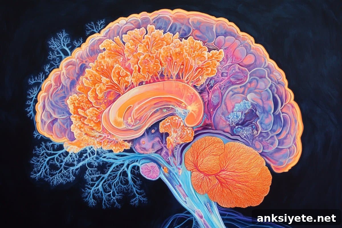
Summary: The brain’s meningeal lymphatic system is essential for clearing waste and supporting immune surveillance, yet how it forms and is positioned has been unclear. New research using zebrafish and high-resolution in vivo imaging reveals that neural activity controls meningeal lymphatic development by regulating a specialized population of glial cells that produce Vegfc, a growth factor crucial for lymphatic formation.
These Vegfc-producing glial cells work closely with ccbe1-expressing fibroblasts that process Vegfc into its active form. Together they guide the growth and precise localization of lymphatic endothelial cells along the brain surface, keeping lymphatic vessels confined to the meninges and preventing them from invading the brain parenchyma. This coordinated interaction highlights how the brain actively shapes its own immune microenvironment.
Key Facts
- Neural activity controls lymphatic development: Increased neuronal stimulation (for example, visual input) enhances meningeal lymphatic growth via Vegfc-expressing glia; reduced activity (for example, visual deprivation) diminishes this development.
- Glia-fibroblast cooperation is essential: slc6a11b+ radial astrocytes secrete Vegfc, but ccbe1+ fibroblasts are required to process pro-Vegfc into mature Vegfc, restricting its activity to the brain-meningeal interface.
- Barrier to immune invasion: This neural-glia-fibroblast axis confines lymphatic endothelial cells to the leptomeninges, preventing lymphatic invasion into brain tissue that could disturb immune homeostasis.
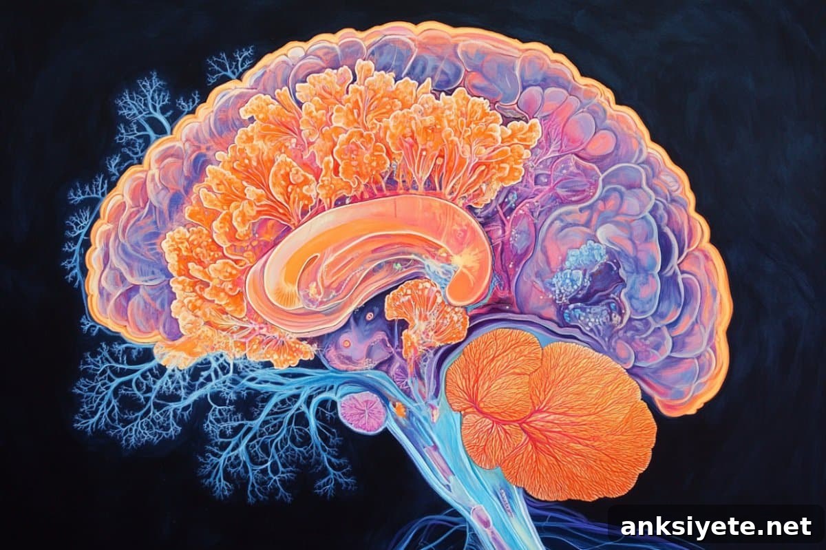
A team led by Dr. Du Jiulin at the Institute of Neuroscience, Center for Excellence in Brain Science and Intelligence Technology, Chinese Academy of Sciences, published these findings in Cell. Their approach combined long-term in vivo imaging in zebrafish with targeted genetic manipulations and experiments that altered neural activity to reveal how meningeal lymphatics form and are patterned.
Using live imaging, the researchers observed that changes in neural activity produce proportional changes in the number and development of mural lymphatic endothelial cells (muLECs) within the leptomeninges. Enhanced sensory input such as visual stimulation increased muLEC formation, while reduced sensory input decreased it. These results indicate that neural function directly influences meningeal lymphatic development.
The study pinpointed Vegfc as a central molecular mediator. A distinct glial subpopulation identified by slc6a11b expression—referred to as slc6a11b+ radial astrocytes (RAs)—expresses Vegfc and extends cellular processes to the brain surface. These slc6a11b+ RAs are the primary source of Vegfc in the brain model studied. Genetic deletion of slc6a11b+ RAs compromised muLEC formation, while enhancing Vegfc signaling in these glia promoted lymphatic development.
Importantly, the researchers demonstrated that neural activity modulates both the activity of slc6a11b+ RAs and their Vegfc expression levels. However, Vegfc secreted by glia is initially released as an inactive precursor (pro-Vegfc). Conversion to the active, mature form (mVegfc) requires the proteolytic processing activity provided by ccbe1-expressing fibroblasts. This intercellular cooperation confines mVegfc to the brain-meningeal interface and thereby restricts lymphatic endothelial cell growth to the brain surface.
By restricting active Vegfc to the meninges, the glia–fibroblast partnership creates a spatially precise signaling environment that prevents muLECs from infiltrating the brain parenchyma. Such invasion could disrupt local immune balance and potentially contribute to neurological dysfunction. The newly described neural-glia-fibroblast-lymphatic axis therefore functions as a protective mechanism that aligns lymphatic architecture with neural activity and regional needs.
This work provides a conceptual framework for how brain function and inter-tissue cellular interactions shape lymphatic development. It suggests that the brain can adapt its lymphatic network in response to changing activity levels, ensuring efficient clearance and immune surveillance where and when it is needed most. Understanding this regulatory axis may open new avenues for exploring the role of meningeal lymphatics in neurological disorders and for designing interventions that target lymphatic function in disease.
About this neuroscience research news
Author: Liu Jia
Source: Chinese Academy of Science
Contact: Liu Jia – Chinese Academy of Science
Image: The image is credited to Neuroscience News
Original Research: Open access.
“Neural-activity-regulated and glia-mediated control of brain lymphatic development” by Du Jiulin et al., Cell. DOI: 10.1016/j.cell.2025.04.008
Abstract
Neural-activity-regulated and glia-mediated control of brain lymphatic development
While the nervous system influences peripheral immune responses, whether and how the brain regulates immune tissue development has been unclear. Meningeal mural lymphatic endothelial cells (muLECs), embedded in the leptomeninges, form an immune niche that supports brain immunosurveillance.
This study reports that the brain controls muLEC development through a specialized glial subpopulation, slc6a11b+ radial astrocytes (RAs), and that this control is modulated by neural activity in zebrafish. slc6a11b+ RAs extend processes to the meninges and govern muLEC formation by expressing vascular endothelial growth factor C (vegfc). Neural activity regulates muLEC development in a manner that depends on Vegfc produced by these glia.
Furthermore, slc6a11b+ RAs cooperate with ccbe1+ fibroblasts to limit muLEC growth to the brain surface by controlling the local distribution of mature Vegfc. Together, these findings reveal a glia-mediated, neural-activity-regulated mechanism for brain lymphatic development and emphasize the importance of coordinated inter-tissue cellular interactions during development.