
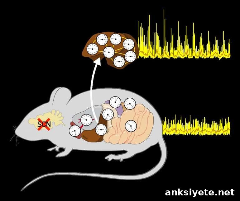
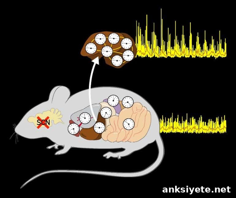
Summary: Using a new imaging technique based on bioluminescence, researchers show that the cellular clocks within a single organ can remain synchronized even when the brain’s master clock and other body clocks are absent.
Source: University of Geneva
Circadian clocks regulate metabolic and physiological processes across roughly 24-hour cycles and are fundamental to life. In humans, disruption of these clocks contributes to metabolic disorders such as diabetes and serious liver disease. Despite decades of study, many basic questions remain about how clocks at the cellular and organ levels are coordinated.
Using an organ-specific bioluminescence monitoring system, a research team at the University of Geneva (UNIGE) demonstrated that the cells composing a single organ can maintain coordinated timing without signals from the brain’s central pacemaker or from other organs. By restoring circadian function only in the liver of otherwise arrhythmic mice, the researchers showed that non-neuronal cells can establish and preserve synchrony on their own.
The full study appears in the journal Genes & Development.
For many years, scientists believed that peripheral circadian rhythms were entirely entrained by a single central clock in the brain. More recently, however, molecular clocks were discovered in nearly every cell type, revealing a distributed system of oscillators. The current work addresses how much the central clock is required to coordinate those peripheral clocks.
“Until now, the prevailing view was that the brain clock was essential for aligning all peripheral clocks,” explains Ueli Schibler, honorary professor at the UNIGE Faculty of Science and initiator of this project. “But the tools commonly available made it difficult to test that idea directly.”
Flore Sinturel, a researcher in the Department of Medicine at UNIGE Faculty of Medicine and the study’s first author, adds: “To test this hypothesis we needed a way to follow circadian gene expression continuously and noninvasively in freely moving animals, for extended periods, both with and without a functional brain clock.”
Bioluminescence to study circadian rhythms
In 2013, Professor Schibler’s lab developed an imaging technology—now commercially available—that enables long-term, organ-specific monitoring of circadian activity. The system relies on a bioluminescent reporter: mice carry a circadian-controlled gene that produces the enzyme luciferase. When luciferin is provided in the drinking water, oxidation by luciferase generates photons. A photomultiplier detects and records the light emitted over time, providing continuous readouts of the reporter gene’s circadian expression in a specific organ.
Liver clock cells remain phase-coupled without external timing cues
When the researchers removed the central brain clock, clocks across different organs fell out of phase with one another. Yet within the liver itself, circadian oscillations remained robust and tightly coordinated. This indicates that while the brain’s master clock can align organs to a common phase, cells within an individual organ can communicate sufficiently to stay synchronized with each other even in the absence of central or external timing cues.
“The assumption that only neurons have sufficiently strong connections to enforce circadian coordination is challenged by these findings,” says Flore Sinturel. “Our results suggest the central clock may be less unique than previously thought.”
To validate this conclusion, the team went further: in mice that were completely arrhythmic—lacking functional clocks throughout the body—they restored clock function exclusively in hepatocytes. The liver then exhibited clear circadian rhythms despite the absence of rhythmicity in other organs. This demonstrates that a clock reinstated in a single organ can operate autonomously and generate coherent rhythms without input from other bodily clocks or environmental cycles.
The investigators are now focused on how hepatocytes maintain phase coherence when isolated from the central pacemaker and external Zeitgebers. Their leading hypothesis is that intercellular coupling—possibly via exchange of signaling molecules or metabolites—allows liver cells to coordinate timing across the tissue.
About this genetics research news
Source: University of Geneva
Contact: Flore Sinturel – University of Geneva
Image: The image is credited to UNIGE
Original Research: Closed access.
“Circadian hepatocyte clocks keep synchrony in the absence of a master pacemaker in the suprachiasmatic nucleus or other extrahepatic clocks” by Flore Sinturel et al. Genes & Development
Abstract
Circadian hepatocyte clocks keep synchrony in the absence of a master pacemaker in the suprachiasmatic nucleus or other extrahepatic clocks
It has long been assumed that the suprachiasmatic nucleus (SCN) acts as the primary synchronizer of peripheral circadian oscillators. Conclusive evidence has been limited, because biochemical time-series measurements are challenging in behaviorally arrhythmic animals. Using long-term bioluminescence recording in freely moving mice, the authors show that the SCN is indeed necessary to keep different organs synchronized with one another. Surprisingly, however, circadian oscillations persist within the livers of mice that lack an SCN or clocks in non-hepatocyte cells. Thus, like SCN neurons, hepatocytes can maintain phase coherence without Zeitgeber input from other organs or environmental cycles.