
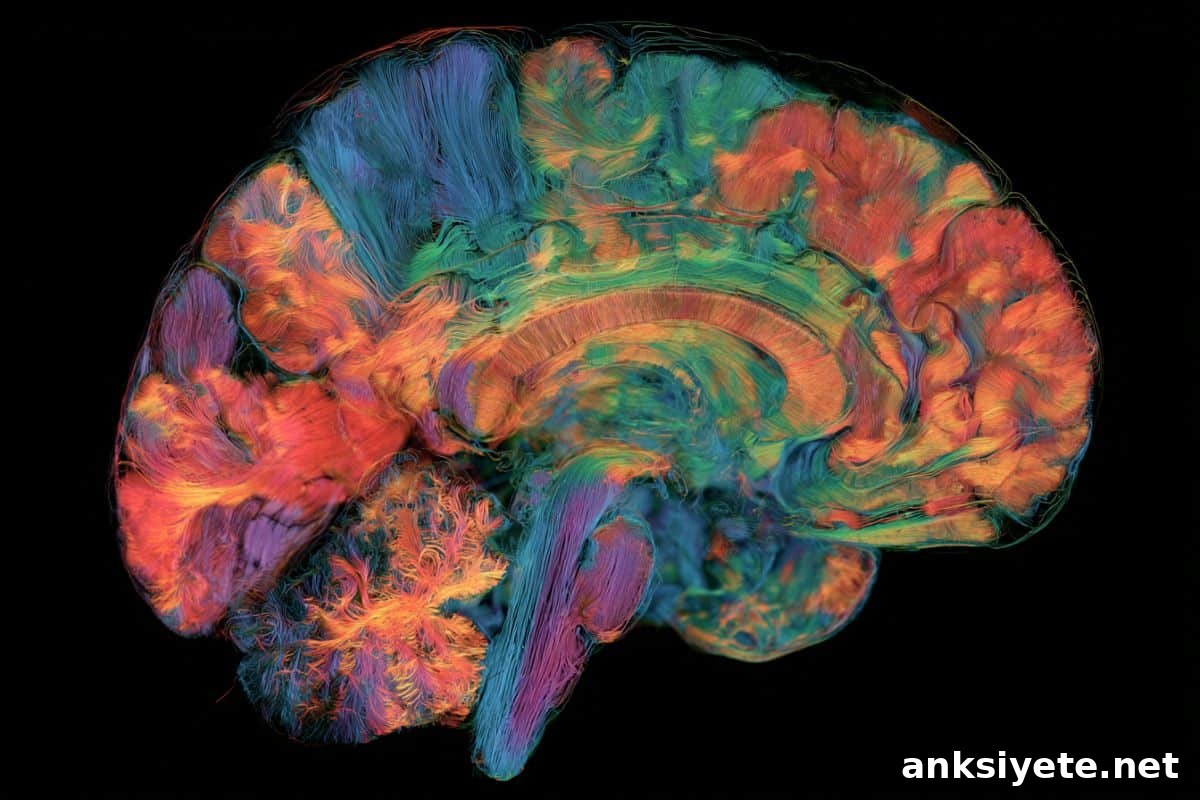
Summary: Researchers trained a deep neural network on magnetic resonance imaging (MRI) scans from nearly 15,000 cognitively healthy adults aged 19 to 100. Moving beyond a single-number “brain age,” the model produces high-resolution 3D maps that estimate local brain age at the voxel level. When applied to people with mild cognitive impairment (MCI) and Alzheimer’s disease (AD), the AI revealed focal patterns of accelerated aging concentrated in the hippocampus, amygdala and frontal–temporal regions, and these localized changes correlated closely with cognitive test performance.
Key Facts
- Voxel-level spatial mapping: Instead of a single global brain-age estimate, the model generates anatomically detailed 3D maps that quantify aging for individual voxels across the entire brain volume.
- Baseline asymmetry and regional trends: In healthy adults, frontal and temporal regions tend to appear biologically older than parietal and occipital regions. The right hemisphere also shows slightly more advanced structural aging than the left, independent of handedness.
- Localized neurodegenerative acceleration: Participants with MCI and AD exhibited pronounced regional age acceleration in structures typically affected early in Alzheimer’s disease, including the hippocampus, amygdala and deep memory-related pathways.
- Structure–function relationship: Higher local brain age was associated with poorer cognitive performance on standardized tests, with the strongest couplings observed in participants with more advanced disease.
- Clinical potential: The approach could enable more precise tracking of regional treatment effects in clinical trials and earlier detection of dementia risk before global measures become abnormal.
Source: USC
Overview
USC researchers led by Associate Professor Andrei Irimia developed a deep-learning method that maps local brain aging across the adult lifespan. Trained on nearly 15,000 high-quality T1-weighted MRI scans from cognitively normal individuals, the model produces three-dimensional heatmaps that show where brain tissue appears older or younger than expected for a person’s chronological age. This spatially detailed view reveals regional patterns of vulnerability and resilience that single-number brain-age metrics can mask.
The model was trained using MRI scans from 14,748 cognitively normal adults, ages 19 to 100, pooled from multiple public datasets. Researchers then applied the model to more than 1,900 additional scans from the Alzheimer’s Disease Neuroimaging Initiative (ADNI), including cognitively normal participants and people diagnosed with mild cognitive impairment and Alzheimer’s disease. The voxel-level estimates of local brain age were compared with cognitive assessments to evaluate structure–function relationships.
Across healthy adults, the model consistently found relatively advanced aging in frontal and temporal lobes—regions involved in decision making, memory and complex cognition—compared with parietal and occipital areas that support sensory processing and spatial awareness. The right hemisphere showed modestly greater structural aging than the left, a pattern that did not depend on whether participants were right- or left-handed.
As cognitive impairment progressed, these regional differences became more pronounced. Compared with cognitively normal adults, people with MCI and AD showed significantly older local brain ages in several cortical and subcortical structures known to be affected early by Alzheimer’s pathology, particularly the hippocampus and amygdala. Importantly, deviations from normative regional aging were associated with worse cognitive performance on tests tapping functions supported by those regions.
Why local brain age matters
Traditional global brain-age biomarkers compress complex neuroanatomy into a single number, which can obscure where the brain is most vulnerable. Estimating local brain age at the voxel level yields anatomically interpretable maps that highlight spatial patterns of aging and degeneration. Such maps can help clarify why some individuals experience selective declines in specific cognitive abilities and may provide a more sensitive tool for detecting early neurodegeneration.
Because the model provides regional readouts, it offers a framework for monitoring whether experimental drugs or interventions slow structural decline in particular brain circuits. That regional specificity could be valuable for precision medicine, trial design and personalized risk assessment, provided the method is validated on diverse clinical and longitudinal datasets.
Limitations and next steps
The authors emphasize that the model is currently a research tool. It was trained primarily on research-grade MRI data and requires further validation with more diverse clinical imaging and longitudinal follow-up to determine whether local brain-age measures can reliably predict individual progression from healthy aging to MCI or AD. Future work will need to test generalizability across scanner types, populations and repeated measures.
Nonetheless, moving beyond a global brain-age summary toward anatomically detailed, voxel-level estimates represents an important advance in neuroimaging biomarkers. By linking regional structural aging to cognitive outcomes, this approach brings greater precision to our understanding of normal and pathological brain aging.
About the study
The study authors include Nikhil N. Chaudhari (first author), Owen M. Vega Huerta, Samayan Bhattacharya, Nahian F. Chowdhury and Andrei Irimia, together with contributors from the Alzheimer’s Disease Neuroimaging Initiative. Funding came from the National Institutes of Health (R01 AG 079957 to Irimia), the Hanson-Thorell Family Research Scholarship Fund, the Center for Undergraduate Research in Viterbi Engineering (CURVE) at USC and anonymous donors.
Key Questions Answered:
A: Earlier methods reduce an entire MRI to a single age estimate, masking regional differences. The USC deep-learning model estimates brain age for each voxel, producing a detailed heatmap that reveals which regions are aging faster or slower than expected for a given chronological age.
A: Even among cognitively normal adults, frontal and temporal lobes typically appear biologically older than occipital and parietal lobes. The right hemisphere also showed modestly more advanced structural aging than the left, regardless of handedness.
A: Not yet. The model remains a research tool pending additional validation across diverse clinical imaging datasets and longitudinal studies to confirm predictive value for individual patient trajectories.
Editorial Notes:
- This article was edited by a Neuroscience News editor.
- Journal paper reviewed in full.
- Additional context was added by staff to clarify methods and implications.
About this AI and brain aging research news
Author: Elizabeth Newcomb
Source: USC
Contact: Elizabeth Newcomb – USC
Image: The image is credited to Neuroscience News
Original research (open access): Deep learning maps local brain aging in relation to cognition across human adulthood, by Nikhil N. Chaudhari, Owen M. Vega Huerta, Samayan Bhattacharya, Nahian F. Chowdhury, Andrei Irimia and the Alzheimer’s Disease Neuroimaging Initiative. Proceedings of the National Academy of Sciences. DOI: 10.1073/pnas.2532233123
Abstract
Deep learning maps local brain aging in relation to cognition across human adulthood
Brain aging, the strongest risk factor for Alzheimer’s disease (AD), varies across cortical regions. Global brain age (GBA) is an imaging-derived summary that reduces structural aging to a single value, potentially obscuring regional patterns of vulnerability that precede AD. This study introduces a deep-learning architecture trained on T1-weighted MRIs from 14,748 cognitively normal participants across multiple sites to estimate local brain age (LBA) at the voxel level. The resulting spatial maps reveal relatively advanced aging in frontal and temporal lobes compared with parietal and occipital regions.
Across diagnostic stages, findings show progressively advanced frontotemporal aging as neurodegeneration progresses from cognitively normal adults (N = 1,102) to mild cognitive impairment (MCI, N = 354) and Alzheimer’s disease (AD, N = 529). Compared to cognitively normal adults, cortical and subcortical structures that typically manifest early AD pathology exhibit significantly older LBAs in both early MCI and AD (P < 0.05). Deviations from normative regional aging are significantly associated with cognitive performance supported by those regions (P < 0.05), linking anatomical aging to functional outcomes. By quantifying regional variations in brain aging, this framework extends global brain-age models and provides anatomically interpretable measures to improve characterization of typical and pathological aging.