Summary:
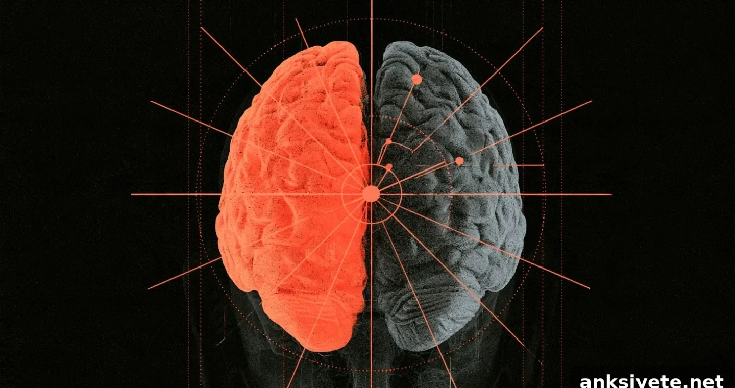
A theoretical paper in Frontiers in Neuroanatomy from researchers at the University of Minnesota offers an evolutionary explanation for why vertebrate brains are cross-wired. The authors argue that contralateral organization—where the left brain controls the right side of the body and vice versa—was not necessarily a set of independent adaptations for touch and movement. Instead, they propose that the optical inversion produced by the eye and the contralateral mapping of the midbrain optic tectum created a structural template more than 500 million years ago that later constrained and guided the evolution of sensory and motor circuits in four-limbed animals.
Key Facts:
- Ancient visual blueprint: Because lenses invert images on the retina, early vertebrates evolved an optic chiasm and a contralaterally mapped optic tectum (the superior colliculus in mammals) to register and orient to stimuli. This arrangement is conserved across all living vertebrates and likely dates back to Cambrian-era ancestors.
- Evolutionary spandrel: The authors suggest many midline-crossing tracts—such as dorsal column, spinothalamic, tectospinal, rubrospinal, and corticospinal pathways—may not have evolved independently for each function but instead followed the pre-existing spatial map set by ancient midbrain vision circuits.
- Co-opted primate dexterity: Although the corticospinal tract originally modulated somatosensory inflow, primates and other dexterous mammals repurposed these contralateral fibers to form direct motor connections with spinal motor neurons, enabling fine finger control.
Source: Frontiers / University of Minnesota
One persistent question in neuroanatomy is why vertebrate nervous systems are contralateral: why does the left cerebral hemisphere monitor and direct the right side of the body, and the right hemisphere do the opposite?
Medical students memorize the many decussations—crossings of tracts in the brainstem and spinal cord—but the evolutionary reasons for this pervasive “cross-wiring” across sensory and motor systems have long been debated.
In a new analysis published in Frontiers in Neuroanatomy, Daniel Wang and Martin Wessendorf, PhD, propose a unifying hypothesis: the brain’s contralateral organization resulted from evolutionary path-dependence rooted in ancient visual systems rather than from multiple independent adaptations.
The optical inversion that started it all
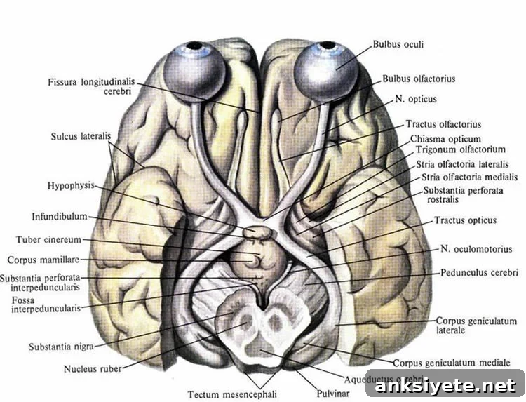
The story begins with vision. A spherical lens projects an inverted image onto the retina, so light from the right visual field lands on the left side of each retina and vice versa. To interpret this inverted input efficiently, early vertebrates evolved an optic chiasm where retinal ganglion cell axons cross the midline so that information from one visual hemifield is routed to the opposite side of the brain.
These retinal projections terminate principally in the optic tectum (the superior colliculus in mammals), an ancient midbrain structure present across vertebrate lineages. The tectum builds a contralateral map of external space used to guide rapid orienting and defensive behaviors. Importantly, in its deeper layers the tectum also receives somatosensory and auditory inputs aligned with the visual map, creating an integrated, contralateral coordinate system.
Because all extant vertebrates possess an optic chiasm and a contralaterally organized tectum, Wang and Wessendorf reason that this architecture was already established in distant Cambrian ancestors.
Conserving the contralateral template in tetrapods
As vertebrates transitioned from water to land and evolved four limbs, new ascending sensory and descending motor pathways emerged to manage limb movement and navigate terrestrial environments. The authors examine major somatosensory and motor tracts and show how they align with the ancient midbrain template.
Two primary ascending somatosensory systems are highlighted:
- Dorsal column–medial lemniscal pathway: Carries discriminative touch, vibration, and proprioception. In tetrapods this pathway ascends the spinal cord and crosses in the medulla; it is conserved across terrestrial vertebrates though absent in many ray-finned fishes.
- Spinothalamic tract: Conveys pain, temperature, and crude touch. This tract crosses the midline within the spinal cord itself before ascending and is found across diverse vertebrate groups, including birds, reptiles, mammals, and cartilaginous fishes.
Descending motor pathways similarly reflect contralateral organization:
- Tectospinal tract: Originating in the tectum’s deep layers, it crosses to the opposite spinal cord to drive reflexive orienting of head and body toward sudden peripheral stimuli.
- Rubrospinal tract: From the midbrain red nucleus, it decussates and projects to the contralateral spinal cord to support coordinated limb movements in tetrapods.
- Corticospinal tract: A later mammalian innovation arising from the neocortex that crosses at the pyramidal decussation. In many mammals it modulates sensory processing in dorsal horn circuits; in primates and other dexterous species these contralateral fibers were recruited to make direct connections with spinal motor neurons, enabling precise, independent finger movements.
Evolution as a pragmatic engineer
Wang and Wessendorf frame evolution as conservative and incremental: rather than redesigning the nervous system from scratch, new circuits tend to be added onto existing scaffolds. Because the optic tectum already coupled contralateral visual maps with somatic inputs to coordinate survival behaviors, later-evolving sensory and motor pathways were constrained to align with that established coordinate system to avoid sensorimotor mismatches.
Under this view, contralateral wiring is best seen as an evolutionary spandrel—an architectural outcome anchored to the optics of the vertebrate eye and the midbrain map it produced—rather than as a collection of separate, optimal solutions for each domain of sensation and action. The authors suggest that future fossil and comparative studies could further trace how this visual blueprint shaped the vertebrate central nervous system.
Editorial Notes:
- This article was edited by a Neuroscience News editor.
- The journal paper was reviewed in full.
- Additional context was added by staff.
About this evolutionary neuroscience research:
- Media contact: Deborah Pirchner
- Source: Frontiers
- Image credit: Image credited to Neuroscience News
- Original research (open access): Frontiers in Neuroanatomy (Oct 1, 2026). “Why is the right side of the brain responsible for the left side of the body? The evolutionary history of a puzzling trait.” Authors: Daniel Delie Wang and Martin W. Wessendorf.
- DOI: 10.3389/fnana.2026.1937941
Abstract
Why is the right side of the brain responsible for the left side of the body? The evolutionary history of a puzzling trait
In humans and most other mammals, the right motor cortex controls spinal motoneurons that move the limbs on the left side of the body. Likewise, the right cerebral cortex predominantly receives sensory information from the left side of the body and the left side of external space. Although many authors—dating back to Ramón y Cajal—have speculated about the adaptive value of contralateral organization, few studies have traced its evolutionary origins.
This paper surveys the principal contralaterally organized circuits in living vertebrates and proposes a novel evolutionary hypothesis. Vision is contralaterally organized in every reported vertebrate: each visual field’s input travels to the contralateral optic tectum (the superior colliculus in mammals) where it is retinotopically mapped. In most tetrapods, somatosensory and auditory pathways are also carried forward by contralateral circuits. Early amniote ancestors appear to have had contralaterally organized motor systems, including tectospinal and rubrospinal tracts. The last common ancestor of mammals likely already possessed a contralateral neocortex with a corticospinal pathway. We conclude that the mammalian neocortex evolved within brains where major motor and sensory circuits were already contralaterally arranged; by adopting this existing organization, the neocortex integrated without incurring substantial “rewiring” costs.