
Summary: A Cell study from Cold Spring Harbor Laboratory reveals a molecular-genetic basis for neuronal cell types. Researchers found that families of genes encoding proteins involved in neurotransmission define neurons by determining their connection partners and modes of communication.
Neuron Types in the Brain Are Defined by Gene Activity That Shapes Their Communication Patterns
Cold Spring Harbor Laboratory (CSHL) researchers report in Cell a major advance: the molecular-genetic rules that distinguish neuronal cell types across the mouse brain appear to be organized around patterns of cell-to-cell communication. By combining high-resolution RNA sequencing with targeted computational analysis, the team identified specific families of genes whose activity determines which cells neurons connect with and how they communicate with those partners.
Neurons form the circuits that underlie perception, thought and behavior. Yet neuroscientists have long struggled to agree on how to define the many distinct neuron types in the brain. The CSHL study led by Professor Z. Josh Huang addresses this challenge by focusing on functionally important gene families that directly shape synaptic communication. The work suggests that a neuron’s communication profile—who it talks to and how—is a defining molecular signature of its type.
Approach and key findings
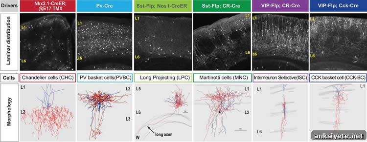
The research team examined six genetically identified types of cortical inhibitory neurons that all use the neurotransmitter GABA. Using a high-resolution RNA sequencing method optimized by first author Anirban Paul, combined with computational tools developed by Assistant Professor Jesse Gillis and colleague Megan Crow, the investigators screened more than 600 gene families for characteristic expression patterns across the six neuron classes.
From this screen, they identified roughly 40 gene families whose expression patterns reliably distinguish among the six inhibitory neuron groups. Strikingly, these families fall into just six functional categories, all of which relate to cell-to-cell communication across the synapse. The gene families include proteins positioned on either side of the synaptic cleft—on membranes facing each other across the narrow gap between a signal-sending (pre-synaptic) neuron and a signal-receiving (post-synaptic) neuron.
According to Huang, the central implication is straightforward: the key genes specify which partners a neuron connects to and how it signals them. In other words, gene expression encodes a neuron’s communication architecture—its synaptic partners and the molecular machinery that mediates their interactions.
Generality and implications
To test whether these communication-related gene families generalize beyond the six GABAergic cell types, the team re-analyzed published datasets from the Allen Brain Atlas. That analysis showed the same six defining gene/protein families help distinguish other neuronal populations, including excitatory neurons, in random samples across the brain. This supports the idea that a core genetic “communication recipe” is broadly conserved and can serve as a principled way to classify diverse neuronal types.
These findings provide an organizing principle that can help resolve longstanding disagreements about neuron classification. While the brain likely contains hundreds or thousands of distinct neuron types, this study suggests classifications grounded in the genes that control synaptic partners and signaling modes reflect biologically meaningful differences rather than arbitrary labels.
Why this matters
By linking specific gene families to the patterns of connectivity and communication that define neuronal identities, this research offers a molecular framework for understanding how diverse neural circuits are constructed. That framework can help neuroscientists sort the complexity of cell types, guide further functional studies, and improve efforts to map brain circuits at cellular resolution.
Funding: This research was supported by the National Institutes of Health, the CSHL Robertson Neuroscience Fund, and the Brain & Behavior Research Foundation/NARSAD.
Source: Peter Tarr — Cold Spring Harbor Laboratory
Image Source: NeuroscienceNews.com images are credited to Huang Lab, CSHL.
Original Research: The study will appear in Cell.