
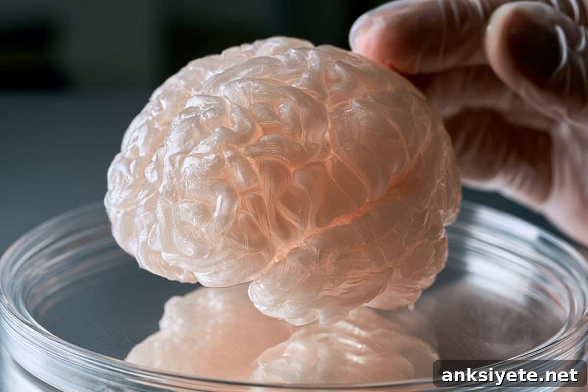
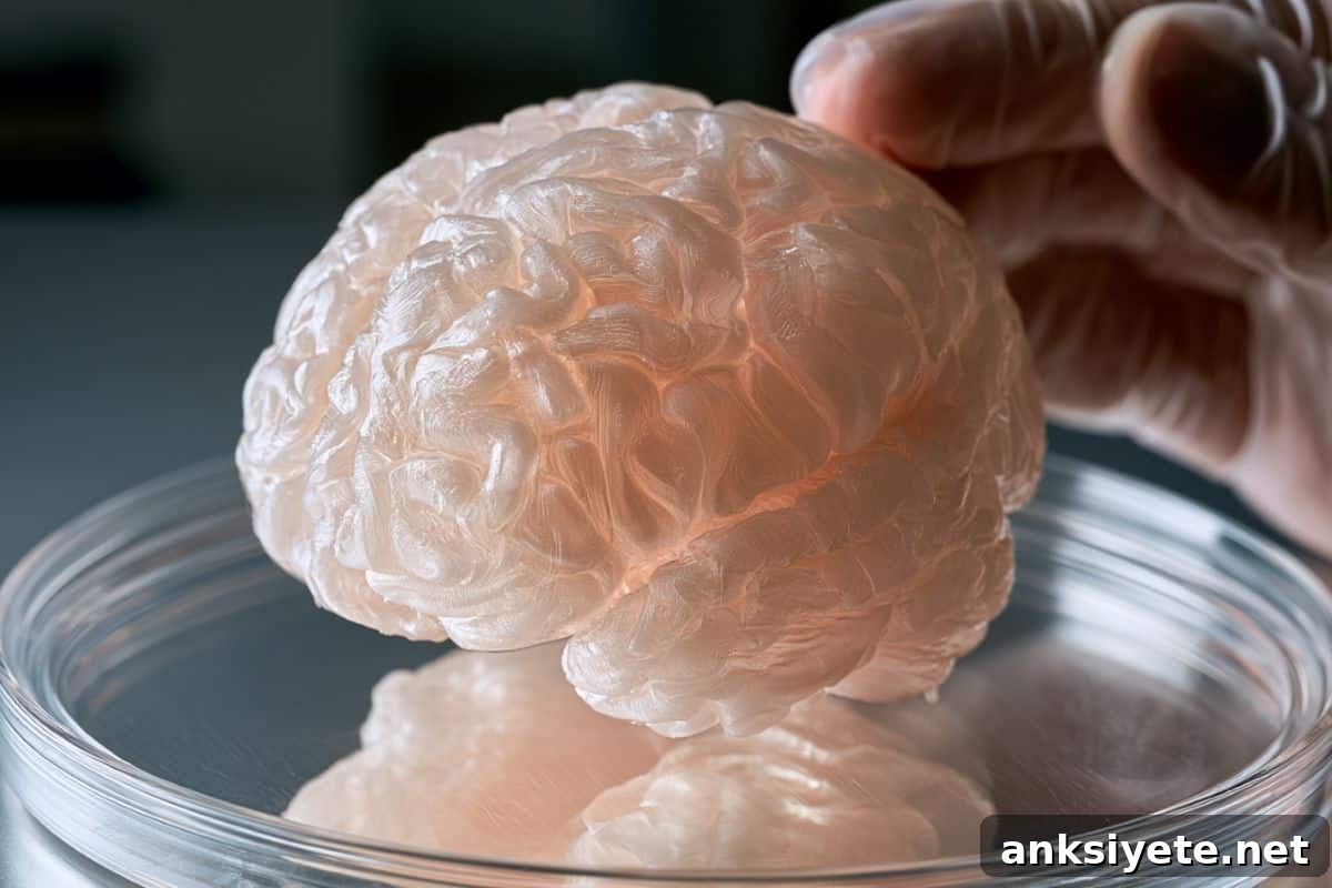
Summary: Researchers have created a highly reproducible three-dimensional human brain tissue model that replicates key neurodegenerative processes seen in Alzheimer’s disease. Developed over nine years from human induced pluripotent stem cells, these self-organizing tissue spheroids combine functional neurons, supportive astrocytes, and immune-competent microglia into compact micro-architectures roughly the size of half a pinhead.
The engineered spheroids express Alzheimer’s-relevant genes and proteins, establish functional synaptic networks, and display active microglial surveillance. The research team validated the platform by inducing hallmark amyloid beta aggregates within the tissues and then clearing those deposits using recently approved anti-amyloid antibodies, demonstrating translational relevance for therapeutic testing.
Key Facts
- Tri-culture cellular interaction: Unlike flat two-dimensional cultures, these 3D spheroids house neurons that form functional synapses, astrocytes that provide metabolic and structural support, and microglia that carry out immune surveillance and clearance functions.
- Rapid self-organization: Under a proprietary differentiation protocol and nutrient regime, the stem-cell–derived cells assemble and self-organize into functional mini-tissue spheroids within about one week.
- Transcriptomic and proteomic fidelity: Extensive molecular profiling confirmed activation of primary genes and proteins associated with Alzheimer’s pathology and normal cell-to-cell signaling in the engineered tissues.
- Validation through plaque clearance: The team induced human amyloid beta deposits in the spheroids and showed that anti-amyloid immunotherapies, acting with active microglia, effectively removed these pathological aggregates.
- Robotic automation and scale-up: The laboratory is adapting the platform to robotic manufacturing to produce thousands of standardized diseased spheroids for industrial-scale drug screening.
Source: LMU
How can Alzheimer’s research be accelerated, improved, and made more predictive of human outcomes? Despite vast global effort and recent therapeutic advances, the disease remains difficult to halt. A major limitation has been the lack of three-dimensional human models that capture the complex cell interactions of diseased brain tissue.
“We still lack 3D models that faithfully recreate the cellular interactions of the human brain in Alzheimer’s disease,” says Dominik Paquet, Professor of Neurobiology at the Institute for Stroke and Dementia Research, LMU University Hospital. His team’s new model addresses this gap and may have broad implications for drug development.
The team published the findings in Nature Neuroscience, summarizing nearly a decade of work to bring this model to functional maturity. “It took us nine years to develop a three-dimensional human brain tissue model that works reliably at all the levels we consider important,” Paquet explains.
For Alzheimer’s research, the functional interactions among cell types and their biochemical activities matter more than reproducing the exact macrostructure of the human brain. The model was therefore designed to prioritize authentic cellular behavior and intercellular communication.
The right recipe for genuine interplay
The model begins with human induced pluripotent stem cells differentiated into neurons, astrocytes, and microglia. The researchers apply a specific combination of signaling factors and culture conditions developed in their laboratory. In an optimized nutrient medium, the differentiated cells adhere and organize themselves, forming compact tissue spheroids about the size of half a pinhead within a week. These spheroids develop core brain-like functions.
Functional and molecular fidelity
Neurons in the spheroids extend processes and form synapses that support network activity. Astrocytes supply metabolic support and maintain homeostasis. Microglia adopt surveillance behaviors and engage in phagocytic clearance of debris and aggregates. Molecular profiling confirmed that genes and proteins central to Alzheimer’s biology are active in the model, supporting its suitability for disease studies.
Reproducible, tunable, and disease-relevant
A key strength of the approach is reproducibility: following the published protocol yields consistent spheroids with uniform composition and function. The system is also manipulable: researchers can induce Alzheimer’s-like pathology, evaluate candidate therapies, and observe microglial responses. The team demonstrated this by inducing amyloid plaque formation and then dissolving those deposits with available anti-amyloid drugs, with microglia clearly participating in the clearance process.
Next step: automation to accelerate drug discovery
To scale the technology for pharmaceutical screening, the group is developing automated production using robotic systems. This will enable manufacture of hundreds or thousands of standardized spheroids that share defined disease features, allowing high-throughput testing of many candidate compounds in a human-relevant cellular setting before advancing to animal studies or clinical trials.
Key Questions Answered:
A: Traditional 2D cultures lack the spatial organization and multicellular interactions of real brain tissue. The 3D spheroid model brings neurons, astrocytes, and microglia together in a structured environment, enabling microglial clearance of debris and pathological aggregates and permitting more realistic cell–cell signaling and network behavior.
A: The model includes three primary cell types derived from human stem cells: neurons that form synaptic connections, astrocytes that support metabolism and structural integrity, and microglia that function as the brain’s resident immune cells and participate in clearing toxic protein aggregates.
A: The system’s reproducibility and planned robotic scaling allow production of large numbers of standardized human tissue models showing Alzheimer’s features. This supports high-throughput screening of compound libraries in a human cellular context, improving the efficiency and predictiveness of preclinical testing.
Editorial Notes:
- This article was edited by a Neuroscience News editor.
- The journal paper was reviewed in full by the editorial team.
- Additional explanatory context was added by staff to clarify technical aspects for readers.
About this Alzheimer’s disease research news
Author: Dominic Anders
Source: LMU
Contact: Dominic Anders – LMU
Image: The image is credited to Neuroscience News
Original Research: Open access. “A reproducible three-dimensional model of human brain tissue to investigate physiological and disease-associated microglia phenotypes” by Julien Klimmt et al., published in Nature Neuroscience. DOI: 10.1038/s43856-026-01767-4
Abstract
A reproducible three-dimensional model of human brain tissue to investigate physiological and disease-associated microglia phenotypes
Stem-cell–based in vitro models hold promise for revealing human brain cell functions and interactions, yet reproducibility, maturation and cell-type diversity have remained barriers. In particular, sustaining mature microglia and modeling neuroinflammation in vitro have been challenging.
The authors report a human induced pluripotent stem cell–derived three-dimensional cortical brain tissue model (3BTM) that incorporates neurons, astrocytes and microglia with high reproducibility, maturity and viability. 3BTMs demonstrate morphological, functional and proteomic maturation across cell types, producing close similarity to their in vivo counterparts.
Integrated microglia persist for more than six months, adopting mature morphologies, functions and gene-expression profiles. When engineered to model Alzheimer’s disease, 3BTMs reproduce essential disease hallmarks, including amyloid deposition, elevated phospho-tau, and neuroinflammatory responses, with microglia shifting toward disease-associated transcriptional states.
Treatment of Alzheimer’s 3BTMs with anti-Aβ immunotherapy cleared deposits and substantially reversed glial disease signatures. Overall, this microglia-containing model provides a human-relevant platform to study physiological and pathological states of brain tissue and to support therapeutic discovery.