
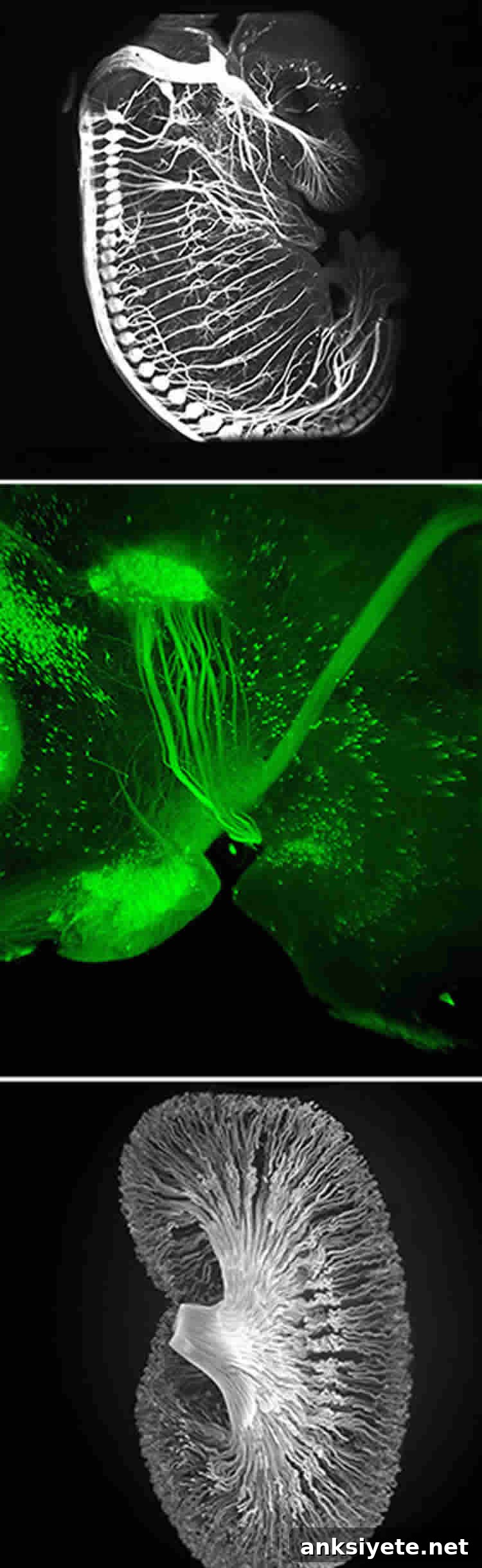
Rockefeller University scientists develop iDISCO: a fast, affordable 3D imaging method for large biological samples
A team of neuroscientists at The Rockefeller University has developed iDISCO, an efficient and cost-effective imaging protocol that enables deep, three-dimensional visualization of molecular structures across large biological tissues. Combining improved immunolabeling with contemporary tissue-clearing approaches and light sheet microscopy, iDISCO makes it possible to label, clear, and image intact organs and large samples with speed and clarity that were previously difficult or costly to achieve. The technique is described in the November 6 issue of the journal Cell.
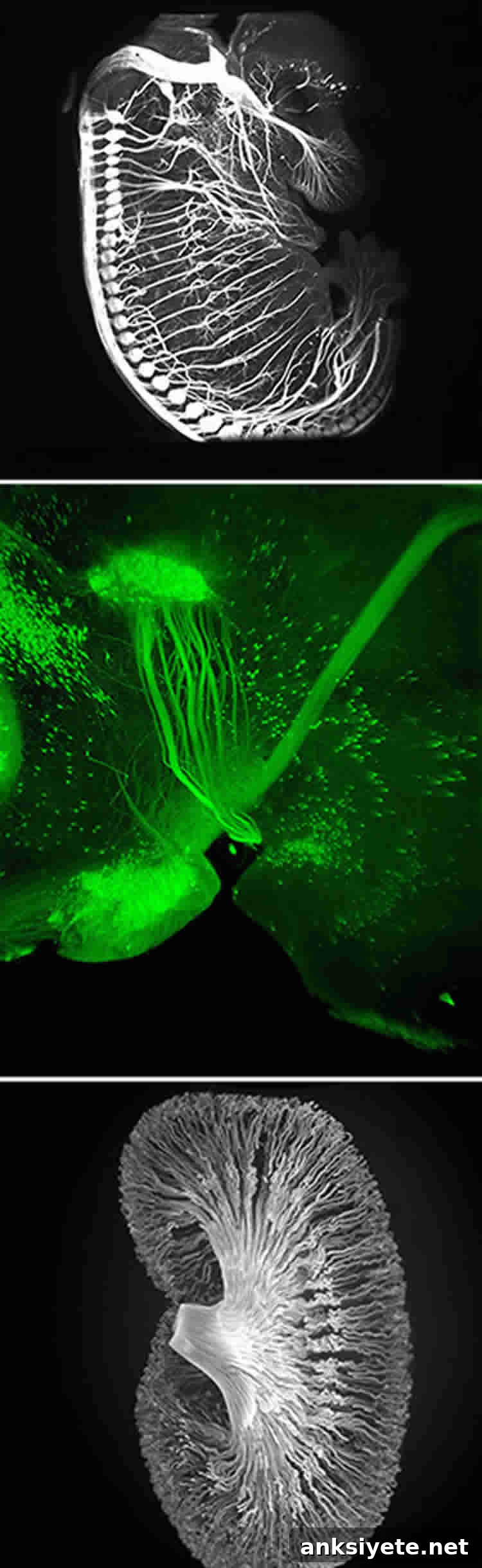
“We optimized multiple parameters of established methods to create a powerful new labeling protocol,” says Marc Tessier-Lavigne, president of Rockefeller University, Carson Family Professor, head of the Laboratory of Brain Development and Repair, and the senior author of the study. “By refining how tissues are labeled, cleared, and imaged, we significantly extended our ability to visualize molecular and cellular architectures deep within intact, complex tissues like the brain.”
iDISCO is presented as an integrated workflow: improved whole‐tissue immunolabeling to tag endogenous molecules, chemical clearing to make tissues transparent, and high-speed light sheet microscopy to capture volumetric data at high resolution. When executed together, these steps enable researchers to examine molecular distributions and anatomical structures in three dimensions across large tissue volumes more rapidly and simply than many earlier methods.
Immunolabeling traditionally has been applied to thin or small samples because antibodies and other probes struggle to penetrate dense tissue. The research team demonstrated that iDISCO can deliver a broad set of commonly used antibodies deep into a variety of tissues and do so more quickly than before. This capability allows scientists to attach detectable tags directly to endogenous proteins and other molecules, complementing experiments that rely on genetic fluorescent reporters.
Clearing is the chemical processing step that renders samples optically transparent so light can travel far into previously opaque tissues. Advances in clearing chemistry have opened new possibilities for volumetric imaging by reducing light scattering and matching refractive indices throughout a sample. The Rockefeller group built on those advances and tuned their labeling and clearing steps to be broadly compatible and reproducible across organs.
Light sheet microscopy provides a practical means to image whole organs or large tissue blocks at cellular resolution while minimizing acquisition time and photodamage. Because the microscope illuminates only a thin plane at a time, it captures high-resolution optical sections that can be reconstructed into a full three-dimensional representation of labeled structures — from long-range neuronal axons to fine epithelial networks in organs like the kidney.
“For neuroscientists, one of the most impactful applications has been tracing axonal pathways in both developing and adult brains,” says Nicolas Renier, a postdoctoral associate in the Tessier-Lavigne laboratory and co-first author of the study. “We were surprised by how clearly we could image these detailed circuits inside the intact adult brain.”
Co-first author Zhuhao Wu adds, “Being able to visualize circuit formation in larger embryos gives us the opportunity to examine nervous system development at stages when structures are more fully formed, opening new directions for developmental studies.”
Beyond neuroscience, iDISCO’s combination of deep, rapid immunolabeling, robust clearing, and scalable volumetric imaging has potential applications across many areas of basic biology. It can help researchers map cellular populations, follow complex molecular patterns through tissues, and study organ architecture in health and disease without resorting to serial sectioning or slow, invasive procedures.
About this research
The Cell paper lists Nicolas Renier and Zhuhao Wu as co–lead authors and Marc Tessier-Lavigne as senior author. Additional coauthors include David J. Simon, Jing Yang (both in the Tessier-Lavigne laboratory), and Pablo Ariel from Rockefeller University’s Bio-Imaging Resource Center. The study is titled “iDISCO: A Simple, Rapid Method to Immunolabel Large Tissue Samples for Volume Imaging” and is available as open access in Cell. DOI: 10.1016/j.cell.2014.10.010. The work was published online October 29, 2014, and appears in the November 6 issue.
Contact: Franklin Hoke, Rockefeller University press office.
Image credit: adapted from Rockefeller University press materials.