
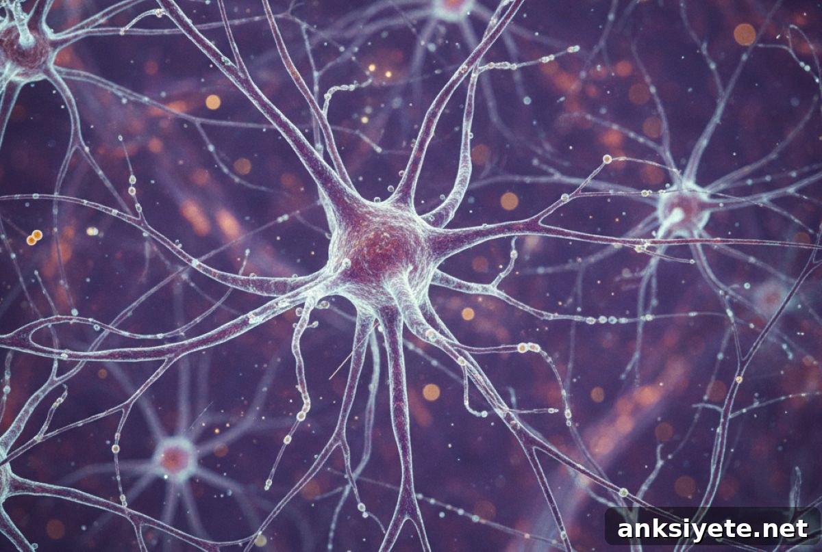
Summary: Researchers have identified a previously unknown cellular barrier in the brain, called the “base barrier cells.” Found at the base of the choroid plexus, these cells form tight molecular junctions that act as a selective gatekeeper, regulating exchanges between the body’s circulation and the brain’s protected environment.
The study shows that this barrier is robust in healthy conditions but becomes compromised during systemic inflammation and severe infection. This discovery reshapes our understanding of brain anatomy and highlights a new target for therapies aimed at preventing or reducing neuroinflammation.
Key Facts
- The “Base Barrier” Discovery: A distinctive population of cells at the base of the choroid plexus forms a secondary, high-security seal that had not been fully recognized previously.
- Vulnerability to Infection: Under systemic inflammatory conditions, the tight junctions that hold these cells together weaken, potentially creating a backdoor for harmful substances and immune cells to enter the central nervous system.
- Human Relevance: These gatekeeper cells are present in both mouse and human tissue, indicating the mechanism is conserved and relevant to human brain health and aging.
Source: VIB
VIB and Ghent University researchers have identified and characterized a previously unknown cellular barrier in the brain, providing new insight into how the brain is protected from the rest of the body.
Published in Nature Neuroscience, the study also describes a pathway through which peripheral immune activity can affect the brain.
Prof. Roosmarijn Vandenbroucke (VIB–UGent Center for Inflammation Research) notes: “These findings reveal how vulnerable and protectable the brain is, opening new possibilities for targeted interventions in brain disorders.”
The brain is shielded by multiple barrier systems that preserve a stable internal environment and prevent harmful substances and pathogens from entering. The blood-brain barrier is the most well-known of these defenses, while the choroid plexus—a small, highly vascularized structure within the brain’s fluid-filled ventricles—produces cerebrospinal fluid (CSF) and forms the blood–CSF barrier.
Despite its critical role, the detailed cellular organization of the choroid plexus and the mechanisms by which it protects the brain have been incompletely understood. The new study fills important gaps in that knowledge.
A new line of defense for the brain
Researchers in the Vandenbroucke lab used advanced single-cell gene expression profiling and high-resolution microscopy to map the cellular landscape of the choroid plexus. They identified a unique cell population at the choroid plexus base, which they named base barrier cells.
These base barrier cells are interconnected by adherens and tight junctions—protein complexes that form a continuous seal. Functionally, this creates compartmentalization between the choroid plexus stroma, the cerebrospinal fluid, and the brain parenchyma, effectively adding an internal, high-security barrier at a strategic location.
“We’ve uncovered a new line of defense for the brain,” says Prof. Vandenbroucke. “These cells form a dynamic gate at a location we had not fully appreciated. That makes this both a fundamental anatomical insight and a promising target for studying brain disease.”
A dynamic barrier vulnerable to disease
The investigators found that the base barrier is not fixed. In healthy animals, it restricts even small molecules from freely passing from the blood-rich choroid plexus into the CSF and adjacent brain tissue. However, during systemic inflammatory responses such as severe infection, the integrity of these junctions is reduced.
Dr. Daan Verhaege, who completed his PhD in the Vandenbroucke lab, explains: “During inflammation, base barrier cells become more permeable, which may allow toxins and immune cells to cross into the central nervous system. This could explain why systemic illnesses can produce neurological symptoms.”
Base barrier cells arise early in development from meningeal mesenchymal precursors and persist throughout life. Their conservation across species and presence in human tissue underline their physiological significance and the potential clinical relevance of the discovery.
By defining this new barrier and its failure points, the study creates avenues to investigate neurological disorders and neuroinflammation further. Therapeutic strategies that strengthen or preserve the base barrier could help prevent harmful peripheral immune influences from reaching the brain and may complement approaches targeting the blood-brain barrier.
Key Questions Answered:
A: The blood–brain barrier remains a primary defense, but this discovery reveals the brain’s protection is layered. The base barrier functions like an internal security gate that reinforces compartmentalization around the brain’s fluid-filled chambers.
A: Severe systemic inflammation can weaken the tight junctions between base barrier cells. When those junctions fail, the gate can open, permitting toxins and inflammatory cells to enter the CSF and potentially reach brain tissue, which helps explain neurological symptoms during systemic illness.
A: Yes. Because base barrier cells act as gatekeepers at the choroid plexus, they represent a potential therapeutic target. Approaches that preserve or reinforce these molecular junctions might reduce neuroinflammation and slow progression of conditions where peripheral immune activity contributes to brain injury.
Editorial Notes:
- This article was edited by a Neuroscience News editor.
- The original journal paper was reviewed in full.
- Additional context was provided by the editorial staff.
About this neuroscience research news
Author: Gunnar De Winter
Source: VIB
Contact: Gunnar De Winter – VIB
Image: Image credit: Neuroscience News
Original Research: Open access. “Base barrier cells provide compartmentalization of choroid plexus, brain and CSF” by Daan Verhaege et al., Nature Neuroscience. DOI: 10.1038/s41593-025-02188-7
Abstract
Base barrier cells provide compartmentalization of choroid plexus, brain and CSF
The choroid plexus (ChP), located in the brain ventricles, is largely composed of epithelial cells that produce cerebrospinal fluid (CSF) and contribute to the blood–CSF barrier. At sites where the ChP attaches to the brain, researchers discovered a distinct population of fibroblast-like cells, termed choroid plexus base barrier cells (BBCs).
The BBCs originate from meningeal mesenchymal precursors, appear early in development, persist throughout life, and are conserved across species. Transcriptionally, BBCs resemble meningeal arachnoid barrier cells and are interconnected by both adherens and tight junctions.
Functional evidence supports that BBCs act as a barrier controlling communication between the periphery and the central nervous system. During inflammatory challenges, loss of barrier integrity and immune cell crossing were observed. Overall, these findings identify a barrier at the ChP base that is essential for compartmentalizing the ChP stroma, brain parenchyma, and CSF, and for protecting the central nervous system.