
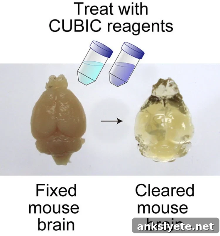
Researchers at the RIKEN Quantitative Biology Center in Japan, working with multiple Japanese institutes, report a straightforward and rapid approach for whole-brain imaging that enables three-dimensional analysis of gene expression patterns and neural circuits at the systems level.
*Please see the notes for important information regarding this research.*
One of the central challenges in systems biology is connecting cellular-scale activity to behavior and function at the level of the whole organism. The brain is a prime focus of this effort, as scientists seek to understand how neuronal activity gives rise to cognition, circadian rhythms and other complex processes. Achieving this goal requires tools that can visualize the entire brain with single-cell resolution and map gene expression, neuronal types and circuit connections in three dimensions.
Traditional approaches to whole-brain imaging typically rely on making fixed tissue highly transparent to minimize light scattering, labeling cells with fluorescent probes, and imaging many optical sections to reconstruct a 3D volume. However, existing clearing and imaging methods have often been limited by long processing times, probe incompatibilities, signal loss, or difficulties scaling from small to large brains. The new method described here, called CUBIC (Clear, Unobstructed Brain Imaging Cocktails and Computational Analysis) and reported in the journal Cell, addresses many of these limitations with a simple, rapid clearing protocol based on aminoalcohols and an imaging pipeline that achieves single-cell resolution across whole brains.
Combined with light sheet fluorescence microscopy, CUBIC was evaluated on several mammalian specimens, including mouse and primate brains, demonstrating that the approach is scalable across different brain sizes. The method generated high-resolution 3D datasets that reveal spatial and temporal patterns of gene expression, for example in hypothalamic regions involved in circadian rhythm regulation. By acquiring image data from multiple directions and computationally merging them, researchers can produce complete, high-quality reconstructions of whole brains and directly compare anatomical or molecular differences across experimental conditions.
CUBIC offers several practical advantages compared with many earlier protocols. The tissue clearing procedure is streamlined: fixed samples are sequentially immersed in only two reagents for relatively short periods, simplifying preparation and improving reproducibility. The reagents are based on aminoalcohol chemistry, which effectively reduces light scattering while preserving tissue structure. Importantly, CUBIC is compatible with a wide range of fluorescent probes and fluorescent proteins because it causes relatively low signal quenching. This compatibility supports multi-color labeling strategies and use of probes that emit at longer wavelengths, both of which enhance imaging depth and contrast for whole-brain datasets.
Reproducibility and scalability are other strengths of the approach. The authors report that CUBIC can be applied consistently across samples and that it scales from small mouse brains to larger mammalian tissues. While other clearing methods have achieved some of these benefits individually, CUBIC integrates rapid clearing, probe compatibility, multi-color imaging potential and computational analysis into a single workflow, making it a practical option for large-scale studies.
Beyond technical improvements, CUBIC expands what is experimentally possible. The method enables three-dimensional mapping of gene expression and neural circuitry at a systems level that was difficult or impossible to obtain previously. Because the pipeline supports high-throughput imaging, it creates opportunities to analyze the localized effects of genome editing on cell populations across entire brains, to map neuronal projections comprehensively, and to compare anatomical or molecular changes induced by environmental conditions or genetic manipulations.
Lead authors and contributors emphasize that this work is a step toward even broader applications. Last author Hiroki Ueda notes optimism about extending the approach: “In the near future, we would like to apply CUBIC technology to whole-body imaging at single cell resolution.” This aspiration highlights the potential for CUBIC-like workflows to inform not only neuroscience research but also systemic analyses of development, disease models and whole-organism responses.
Contact: Jens Wilkinson – RIKEN
Source: RIKEN press release
Image Source: The image is credited to Susaki et al/RIKEN/Cell and is adapted from the RIKEN press release
Original Research: Abstract for “Whole-brain imaging with single-cell resolution using chemical cocktails and computational analysis” by Etsuo A. Susaki, Kazuki Tainaka, Dimitri Perrin, Fumiaki Kishino, Takehiro Tawara, Tomonobu M. Watanabe, Chihiro Yokoyama, Hirotaka Onoe, Megumi Eguchi, Shun Yamaguchi, Takaya Abe, Hiroshi Kiyonari, Yoshihiro Shimizu, Atsushi Miyawaki, Hideo Yokota and Hiroki R. Ueda. Published in Cell. DOI: 10.1016/j.cell.2014.03.042. Publication stage: In Press Corrected Proof
Important disclaimer: The abstract page includes this notice: “Note to users: Uncorrected proofs are Articles in Press that have been copy edited and formatted, but have not been finalized yet. They still need to be proof-read and corrected by the author(s) and the text could still change before final publication.”