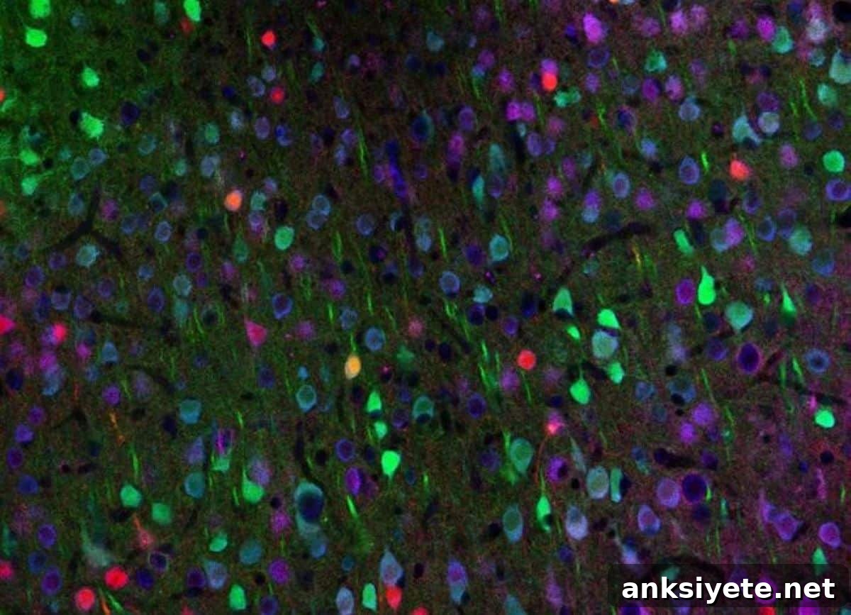
Summary: By tracking living circuit dynamics in transgenic mouse models, researchers uncovered a pronounced, previously hidden imbalance in the motor cortex: a specific class of cortical inhibitory neurons—vasoactive intestinal peptide (VIP) neurons—becomes markedly underactive and near-silent as Huntington’s disease progresses.
Using optogenetics, a precise light-based method, the team reactivated these suppressed neurons. That targeted stimulation reopened a window for neuroplasticity, restored healthier brainwave patterns, reversed motor learning deficits and produced behavioral improvements that lasted after stimulation ended.
Key Facts
- Intercellular imbalance: Contrary to longstanding assumptions that inhibitory cortical cells are spared in Huntington’s disease, this study reveals a large-scale imbalance in inhibitory networks—some interneuron types become hyperactive while others become nearly silent.
- VIP neurons are silenced: Vasoactive intestinal peptide (VIP) interneurons show a pronounced drop in activity. Because VIP neurons help enable cortical reorganization and learning-related plasticity, their silence effectively prevents the diseased brain from adapting and refining circuits during learning.
- Optogenetic reactivation: The team used optogenetics to deliver light-sensitive proteins to VIP neurons and then applied precise light stimulation to override the disease-induced silence in these cells.
- Enduring motor and learning gains: Stimulating VIP neurons gradually normalized activity across the motor cortex and produced rapid, substantial improvements in mice’ ability to learn and perform complex motor tasks.
- Lasting plasticity: Behavioral and physiological gains persisted for days after stimulation stopped, indicating that activation induced long-lasting circuit changes rather than only short-term symptom masking.
- Roadmap to non-invasive approaches: While direct optogenetic brain stimulation is not yet ready for clinical use in humans, the findings identify a clear cellular target and suggest future non-invasive strategies—such as focused electromagnetic or ultrasonic methods—to retune specific, silent circuits from outside the skull.
Source: UCSD
Huntington’s disease is a progressive neurodegenerative disorder in which the degeneration of nerve cells leads to worsening movement and cognitive impairments. Although the genetic mutation—the expanded CAG repeat in the Huntingtin (HTT) gene—is well established, how that mutation disrupts particular cortical circuits and drives behavioral symptoms has remained incompletely understood. This knowledge gap has hindered development of effective therapies.
Researchers at the University of California San Diego, collaborating with colleagues in Germany, used longitudinal in vivo imaging in transgenic mouse models to identify the circuit-level changes that accompany Huntington’s disease progression. They then tested whether restoring activity in a specific interneuron subtype could reverse functional deficits.
“This work shows that correcting defined imbalances in brain circuits can restore function even in a complex neurodegenerative disorder, and highlights the potential of targeting specific cell types to promote recovery,” said Takaki Komiyama, professor in the UC San Diego Departments of Neurobiology and Neurosciences and senior author of the study.
The study was published July 1, 2026, in Nature.
Huntington’s disease arises from an inherited expansion of CAG repeats in the HTT gene, which gradually impairs motor skills and cognition. To map the cellular changes that lead to these impairments, the research team—led by Assistant Project Scientist Sonja Blumenstock—studied how distinct neuron types in the motor cortex respond as disease progresses in transgenic mouse lines carrying the human HTT mutation.
Using advanced two-photon calcium imaging, the investigators followed activity in excitatory projection neurons and three classes of cortical inhibitory interneurons across disease stages. Working with Irina Dudanova’s laboratory in Germany, they discovered that Huntington’s disease disrupts the balance of activity across these cell types: some interneurons become overly active while others, notably VIP interneurons, become profoundly hypoactive.
“Cortical inhibitory cells have received little attention in Huntington’s disease, as for a long time they were considered to be spared from neurodegeneration,” said Dudanova. “Surprisingly, we observed major activity changes—some cell classes are overactive and others nearly silent.”
Previous work from Komiyama’s group had shown that VIP interneurons are important for learning because they enable the brain to enter states that support circuit refinement. Based on that insight, the researchers tested whether artificially restoring VIP activity could re-establish plasticity and improve behavior. They used optogenetics to selectively stimulate VIP neurons in affected mice and then assessed cortical activity and motor learning performance.
Activating VIP neurons restored more normal activity patterns in both VIP cells and their downstream targets, and led to marked improvements in motor learning. Importantly, these behavioral gains persisted for days after stimulation ceased, indicating the intervention induced lasting circuit-level changes rather than only temporary effects.
The findings position VIP interneurons as a vulnerable node in Huntington’s disease and as a promising therapeutic target. The authors propose that modulating VIP neurons opens a “gate” that permits learning-related cortical plasticity to occur despite the underlying genetic defect.
Although optogenetic stimulation as applied in this study is not yet clinically feasible in humans, the work offers a precise cellular target for future non-invasive interventions. Komiyama and colleagues envision developing external, focused stimulation technologies—such as refined magnetic or ultrasonic approaches—that could selectively reactivate silent cell populations without surgery.
“Despite the genetic defect, a focused intervention into circuit activity can produce significant improvements in motor symptoms,” said Dudanova. “If we know which cells to target, we can retune aberrant brain activity patterns. That gives real hope for future therapies.”
More broadly, the study demonstrates that correcting specific circuit imbalances can restore function in a complex neurodegenerative condition, and it suggests similar strategies may be applicable to other disorders that disrupt brain network dynamics.
“We have found a way to allow the diseased brain to learn more effectively,” Komiyama said. “The approach improves behavior in affected mice, and our hope is that related strategies will eventually benefit people with impaired learning and motor function.”
Key Questions Answered:
A: The study shows that the widespread genetic mutation produces specific functional bottlenecks in circuit dynamics. In the motor cortex, the mutation selectively silences VIP interneurons, which act as gatekeepers for learning-related plasticity. Restoring activity in this single cell class allowed broader network function to recover, enabling improved motor behavior despite the persistent genetic defect.
A: Optogenetics is a technique that delivers light-sensitive proteins to specific neurons so those cells can be precisely activated or inhibited with light. Its cellular specificity allowed the researchers to target only VIP neurons, proving that reactivating this particular interneuron type is sufficient to restore motor learning in the mouse models.
A: The study provides a clear cellular target and a mechanistic blueprint. Clinical approaches that non-invasively stimulate or modulate specific cell populations—such as advanced transcranial magnetic or focused ultrasound stimulation—could be developed to mimic the beneficial effects seen in mice without the need for direct optogenetic intervention.
Editorial Notes:
- This article was edited by a Neuroscience News editor.
- The journal paper was reviewed in full.
- Additional context was added by the editorial staff.
About this Huntington’s disease research news
Author: Mario Aguilera
Source: UCSD
Contact: Mario Aguilera – UCSD
Image: Image credit: Komiyama Lab, UC San Diego
Original Research: Open access. “Restoring cortical disinhibition improves Huntington’s disease phenotypes” by Sonja Blumenstock, David Arakelyan, Nicholas del Grosso, Sonja Schneider, Yufeng Shao, Enida Gjoni, Rüdiger Klein, Irina Dudanova & Takaki Komiyama. Nature. DOI: 10.1038/s41586-026-10671-9
Abstract
Restoring cortical disinhibition improves Huntington’s disease phenotypes
Huntington’s disease (HD) is a progressive movement disorder with no cure. Although the monogenic cause of HD is well defined, downstream circuit mechanisms that produce behavioral symptoms are incompletely understood. Cortical dysfunction contributes to HD, but the roles of defined cortical neuronal subtypes in symptom expression have been largely unexplored.
Using longitudinal in vivo two-photon calcium imaging, the authors examined activity in three cortical inhibitory neuron subtypes and in excitatory corticostriatal projection neurons within the motor cortex of the transgenic R6/2 HD mouse model across disease progression.
They observed neuron subtype–specific abnormalities in movement-related activity accompanying motor deficits, including pronounced hypoactivity of VIP interneurons and corticostriatal projection neurons. Similar VIP hypoactivity was also detected in the knock-in zQ175DN HD mouse model. Optogenetic activation of VIP interneurons in R6/2 mice restored healthy activity in VIP cells and their downstream targets and improved motor behavior; these behavioral improvements persisted for days after stimulation. The findings identify cortical interneurons as a potential therapeutic target for Huntington’s disease.