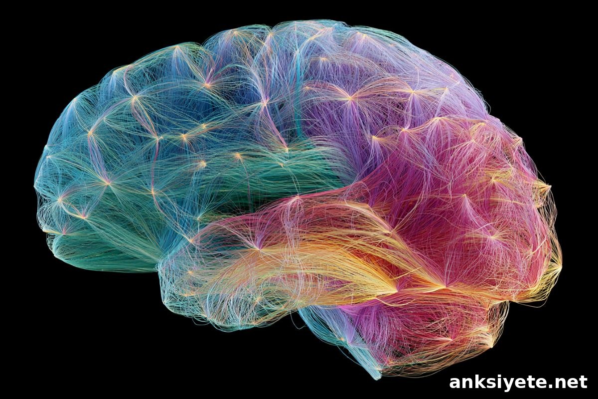
Summary: New research using simultaneous EEG and fMRI demonstrates that the human brain does not run a single unified process. Instead, the connectome coordinates multiple independent, asynchronous streams of information processing that run in parallel. This finding alters how researchers interpret fMRI and EEG signals and opens new possibilities for clinical neuro-diagnostics and studies of cognitive complexity.
Key Facts
- The single-process idea overturned: Concurrent EEG-fMRI recordings show the brain runs several distinct, coordinated processing streams at once, rather than a single process seen at different speeds by each modality.
- Language as an analogy: The brain processes speech on parallel tracks—tracking rapid phonetic cues, assembling words at intermediate speed, and following the broader meaning more slowly—each stream operating independently but together creating comprehension.
- Shared spatial patterns, asynchronous timing: Separate processing streams use the same anatomical networks and similar spatial patterns, and they often activate in the same order — yet each stream unfolds on its own timeline.
- Technical breakthrough: Recording artifact-free EEG inside a powerful, vibrating MRI environment required years of safety work and advanced artifact-cleaning algorithms to preserve true neural signals.
- EEG’s independent clinical value: The study shows EEG captures unique connectome information not reducible to fMRI. This validates using standalone EEG for diagnosis and monitoring, especially where MRI is unavailable, unsuitable, or unaffordable.
- Clinical and translational potential: Revealing parallel asynchronous streams provides a new framework for studying psychiatric, autoimmune, and neurodegenerative conditions—offering ways to track how timing networks are disrupted by dementia, aging, HIV, and other disorders.
Source: Beckman Institute
Postdoctoral researcher Suhnyoung Jun has long been fascinated by how brains work.
Jun is co-first author on a recent paper that investigates the connectome—the brain’s comprehensive network of neural connections—while working in the CONNECTlab with psychology professor Sepideh Sadaghiani.
Using simultaneous electroencephalogram (EEG) and functional magnetic resonance imaging (fMRI) at the Beckman Institute’s Biomedical Imaging Center, Jun and colleagues examined how connectome dynamics unfold across different timescales by capturing both modalities at the same time.
fMRI measures blood-oxygen-level changes and therefore reflects activity at slower timescales; EEG measures electrical activity at millisecond precision. For years many researchers assumed both methods reveal the same underlying brain activity, with fMRI acting as a time-smoothed version of EEG. Jun and her team set out to test that assumption.
Recording EEG and fMRI concurrently allowed the team to compare these signals directly. Their data show the brain operates multiple separate but coordinated processing streams in parallel. Each stream has a distinct temporal profile and unfolds independently, even while using the same spatial pathways.
“It’s like language processing,” Jun explained: “the brain simultaneously tracks the rapid flicker of sounds, the slower arrival of words, and the broader narrative of meaning—each on its own stream.”
Access to the Beckman Institute’s specialized facilities and technical staff was essential. The project spanned nearly five years, including extensive safety training and methodological development. The team overcame major technical obstacles to clean concurrent EEG-fMRI recordings and produced reliable, artifact-minimized datasets thanks to contributions from the Biomedical Imaging Center staff and CONNECTlab researchers.
The study drew on several datasets for validation. Co-first author Thomas Alderson received a Faculty Early Career Development grant from the National Science Foundation. The team combined concurrent EEG-fMRI data from collaborators and a large EEG dataset of 443 participants from the Minnesota Center for Twin and Family Research to strengthen and replicate their findings.
Their results were consistent: multiple asynchronous streams are present across timescales, yet they tend to follow the same spatial blueprints and the same sequences of network activation. In other words, the connectome supports parallel dynamic streams that integrate through shared spatial and temporal organization without reducing to a single process.
Beyond theoretical implications, the findings have practical significance. Demonstrating that EEG provides independent and high-fidelity connectome information supports its use in clinical contexts where MRI is impractical or unavailable. This can help reduce biases in brain-health models that currently rely heavily on fMRI data and exclude populations who cannot undergo MRI scanning.
Jun emphasized the translational promise: “I hope this research helps more patients and informs work that moves toward clinical applications.” The approach offers new ways to study how timing disruptions in the connectome relate to neurological and psychiatric disorders, aging, and autoimmune conditions.
Key Questions Answered:
A: The main limitation was technological. Simultaneous clean recordings were difficult to obtain: fMRI is slow and measures blood flow, while EEG is fast and measures electrical activity. Without a reliable way to record both at the same time, researchers assumed fMRI was simply a slower reflection of EEG. By developing safe, artifact-resistant concurrent recording methods, the Beckman Institute team demonstrated these modalities capture distinct, independent processes.
A: Imagine an orchestra where different sections read the same score but begin at different moments and play at different tempos. The brain’s physical pathways and the sequence in which areas activate are consistent across streams, yet each stream runs on its own timeline and serves different layers of processing.
A: Many clinical and research models rely heavily on expensive fMRI data, excluding individuals who cannot undergo MRI for safety, cost, or access reasons. This study shows EEG carries independent connectome information, enabling clinicians to use lower-cost, portable EEG for diagnosis and monitoring in underserved populations, thereby making brain health assessment more inclusive.
Editorial Notes:
- This article was edited by a Neuroscience News editor.
- The journal paper was reviewed in full for accuracy.
- Additional context was added by the editorial staff to clarify methods and implications.
About this connectome research news
Author: Alejandra Pires
Source: Beckman Institute
Contact: Alejandra Pires – Beckman Institute
Image: The image is credited to Neuroscience News
Original Research: Open access.
“Shared spatial and temporal principles govern connectome dynamics across timescales” by Anne-Lise Giraud, Jeremy Harper, Jonathan Wirsich, Maximillian Kirichenko Egan, Parham Mostame, Samar Wagih ElSayed, Sanmi Koyejo, Sepideh Sadaghiani, Sophia A. Giakas, Stephen M. Malone, Suhnyoung Jun, Thomas H. Alderson, William G. Iacono. PNAS
DOI: 10.1073/pnas.2535464123
Abstract
Shared spatial and temporal principles govern connectome dynamics across timescales
The brain processes information across a wide range of speeds, but how the functional connectome supports multiple concurrent timescales has been unclear. Traditionally, fMRI and electrophysiological methods have been used to study slow and fast dynamics, respectively, and many assumed these methods reflect the same underlying processes filtered differently by each modality’s temporal resolution.
Using simultaneous human fMRI and source-localized EEG, the authors examined instantaneous coactivation patterns—the building blocks of connectome dynamics—across six timescales, from infraslow activity to gamma-band oscillations. They identified streams of recurrent coactivation patterns, or states, that operate in parallel and asynchronously across these timescales, providing a spatial principle that spans speeds. The states also appeared in highly similar sequences at all timescales, revealing a temporal principle that likewise spans speeds.
These results indicate the connectome is composed of multiple dynamic streams running in parallel at distinct speeds, from tens of milliseconds to seconds, rather than a single stream merely filtered by measurement modality. Shared spatial and temporal principles enable the integration of these streams into a unified system. Consequently, studies of human behavior and mental disorders should account for the full range of connectome timescales.