
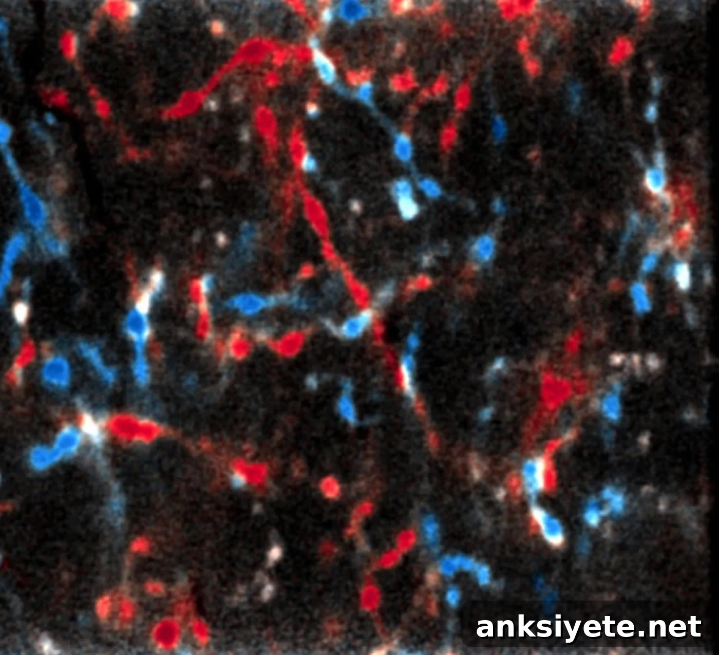
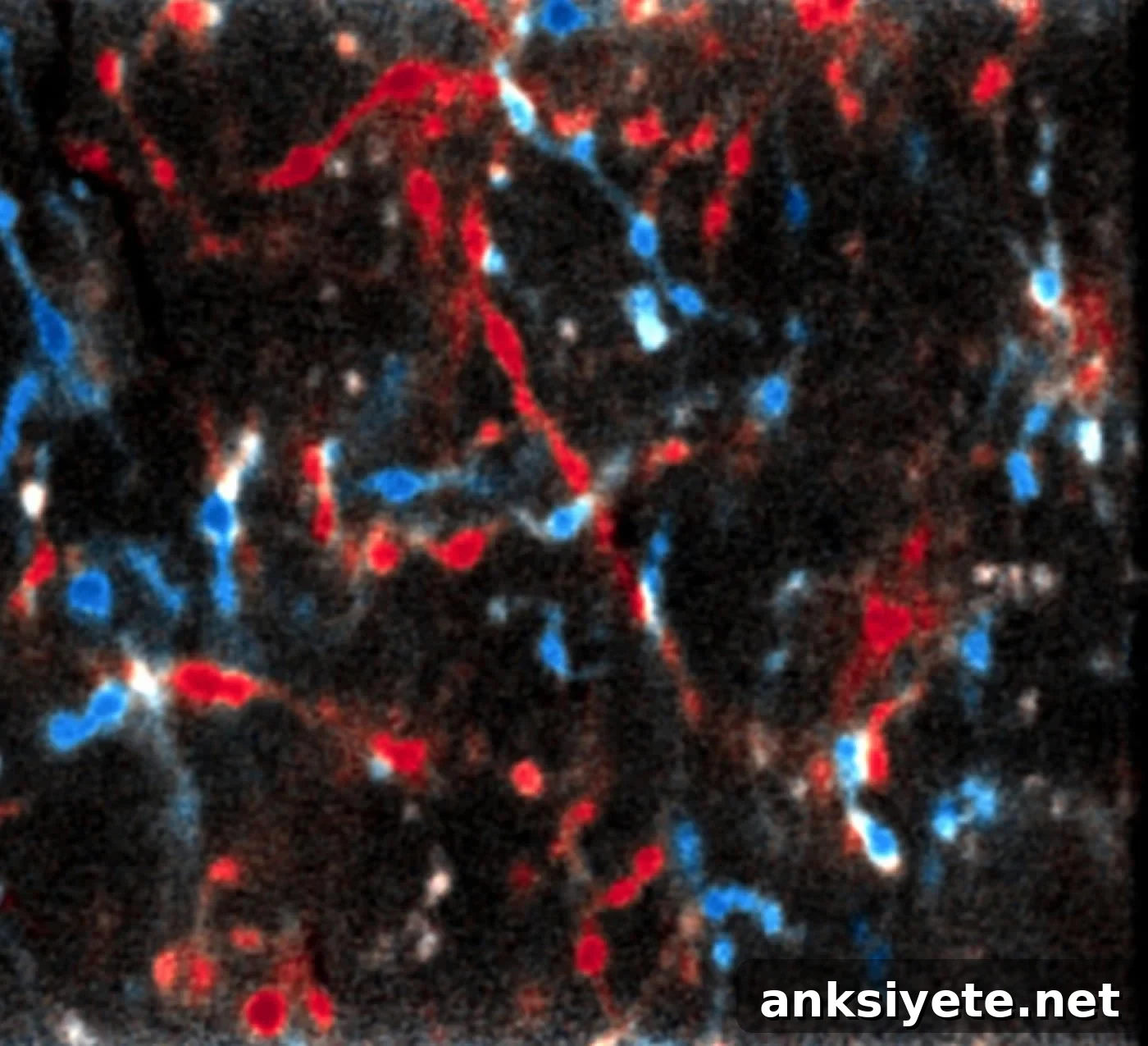
Summary: New findings from researchers at the Max Planck Institute challenge the longstanding view of the thalamus as a passive relay. The study shows the thalamus actively changes its visual responses during learning-related adaptation, indicating a more dynamic role in sensory processing and plasticity than previously believed.
Source: Max Planck Institute.
The cerebral cortex is classically identified as the seat of learning, perception, memory, and conscious behavior. Textbooks typically describe upstream brain regions such as the thalamus as primarily relays that forward sensory input to the cortex and filter signals when necessary. New research from the Max Planck Institute of Neurobiology indicates this account needs revision. In the mouse brain, the thalamus appears to participate actively in visual processing and in experience-dependent plasticity, rather than acting solely as a passive conduit to the cortex.
During early development the brain must learn to interpret and combine input from both eyes to form a coherent visual representation. Neurons in the visual cortex refine their connectivity through experience, enabling efficient processing of visual stimuli. If the signals from each eye are mismatched—such as in cases of strabismus—this can lead to incorrect cortical wiring and reduced visual function. Clinically, temporarily covering the dominant eye during a critical developmental window can encourage the weaker eye’s input to strengthen in the cortex, restoring more balanced visual processing. These well-characterized shifts in the visual cortex have long served as a model for studying cortical learning mechanisms using mouse experiments.
Researchers in Tobias Bonhoeffer’s group investigated neural activity not only in the cortex but also in upstream structures, particularly the thalamus, during monocular deprivation (temporary eye closure). To their surprise, thalamic cells did more than passively forward signals: they altered their responses in response to the change in visual input. “This was completely unexpected, as it has been believed for over 50 years that the thalamus only forwards information and is not actively involved in learning processes,” says Tobias Rose, the study leader.
Previous studies had not detected comparable changes in the thalamus. The authors suggest two possibilities: either the mouse thalamus exhibits species-specific plasticity not seen in other mammals, or earlier experimental methods lacked the sensitivity to detect these thalamic changes. In either case, the new data support a more active role for the thalamus in experience-dependent plasticity.
Additional experiments indicated that the observed changes in thalamic activity are not merely a downstream consequence of cortical adjustments. The thalamic shifts persisted even when cortical activity was suppressed pharmacologically, implying that thalamic neurons can change their eye-specific responsiveness independently of cortical feedback. “It seems we understood less than we thought about how sensory learning is distributed across brain regions. We will need to rethink the thalamus’s contribution to learning,” says Tobias Bonhoeffer.
Source: Stefanie Merker – Max Planck Institute
Publisher: Organized by Neuroscience News.
Image Source: Image credited to MPI of Neurobiology / T. Rose.
Original Research: Abstract for “Lateral geniculate neurons projecting to primary visual cortex show ocular dominance plasticity in adult mice” by Juliane Jaepel, Mark Hübener, Tobias Bonhoeffer & Tobias Rose, published in Nature Neuroscience (published online November 13, 2017).
Max Planck Institute. “The Thalamus Helps the Cerebrum With Learning.” Neuroscience News. Published November 14, 2017.
Abstract
Lateral geniculate neurons projecting to primary visual cortex show ocular dominance plasticity in adult mice
Experience-dependent plasticity in the mature visual system is widely considered to be cortical. Using chronic two-photon Ca2+ imaging of thalamic afferents in layer 1 of binocular visual cortex, the authors provide evidence that challenges this assumption: dorsal lateral geniculate nucleus (dLGN) cells exhibited pronounced ocular dominance (OD) shifts after monocular deprivation in adult mice. While most (86%) dLGN boutons were monocular during normal visual experience, deprivation caused boutons dominated by the closed eye to reduce or lose responsiveness to that eye and often become responsive to the open eye. These changes cannot be fully explained by eye-specific cortical alterations feeding back to the dLGN, since the OD shift in dLGN responses largely resisted cortical inactivation using the GABAA receptor agonist muscimol. The data indicate that OD shifts observed in the adult binocular visual cortex may at least partially reflect plasticity of eye-specific inputs onto dLGN neurons.
“Lateral geniculate neurons projecting to primary visual cortex show ocular dominance plasticity in adult mice” by Juliane Jaepel, Mark Hübener, Tobias Bonhoeffer & Tobias Rose. Nature Neuroscience. Published online November 13, 2017. doi:10.1038/s41593-017-0021-0