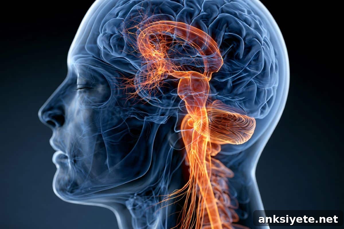
Summary: Researchers show that the effectiveness of anti-CTLA-4 immunotherapy against brain tumors depends not only on local T cells but critically on a robust B-cell and antibody response that is initiated far from the tumor, within the deep cervical lymph nodes of the neck.
By revealing that anti-CTLA-4 stimulates the production of tumor-targeting Immunoglobulin G (IgG) antibodies in tumor-draining deep cervical lymph nodes, this work changes fundamental assumptions in neuro-oncology and suggests a new strategy for treating difficult brain cancers.
Key Facts
- Rethinking the T-cell-centric view: Traditionally, cancer immunotherapy has focused on T cells as the primary effectors. In mouse glioma models, anti-CTLA-4 produced dramatic tumor shrinkage and extended survival, but these benefits disappeared in animals lacking B cells. This finding establishes B cells as essential mediators of anti-CTLA-4 efficacy in these models.
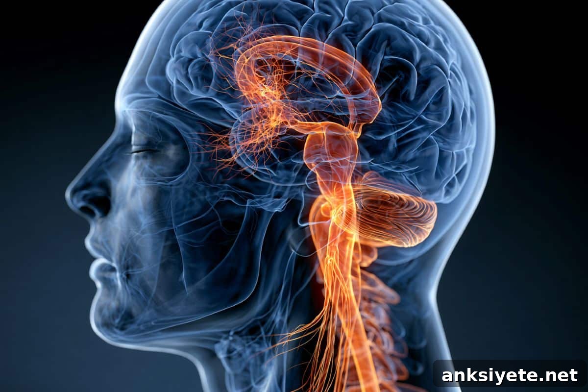
- Deep cervical lymph nodes as the immune hub: The critical B-cell activity did not occur primarily within the brain. Instead, it was concentrated in the deep cervical lymph nodes—neck lymphatic structures that filter cerebrospinal fluid draining from the skull and serve as a major site of immune activation for central nervous system antigens.
- Germinal center activation and IgG production: After anti-CTLA-4 treatment, researchers observed a surge of germinal center B cells working with T follicular helper cells in these lymph nodes. This coordinated response drove an increase in high-affinity IgG antibodies capable of recognizing tumor cells.
- Antibody-mediated opsonization and macrophage clearance: The IgG antibodies trafficked from the neck to the brain and bound to glioma cell surfaces. Bound antibodies flagged tumor cells for macrophages, promoting enhanced phagocytosis and removal of cancer cells by these innate immune effectors.
- Direct in vivo visualization: The team used a dual-reporter glioma model expressing mCherry (red) and EGFP (green) fluorescent proteins and high-resolution intravital imaging to directly observe tumor-infiltrating phagocytes engulfing fluorescent tumor cells after anti-CTLA-4 treatment.
- Implications for cancer immunotherapy: This study provides the first functional evidence that B-cell–driven responses in tumor-draining lymph nodes can determine the success of brain tumor immunotherapy. It expands the conceptual framework beyond local, intratumoral T-cell responses to include distal systemic immune sites as critical contributors to therapeutic outcome.
Source: KAIST
Overview: Researchers investigated why immune checkpoint inhibitors, which release inhibitory signals on immune cells, often have limited benefit for some brain tumors. Their findings point to a crucial role for B cells and antibody responses initiated in tumor-draining lymph nodes as a key component of anti-CTLA-4–mediated antitumor activity.
On July 19, KAIST announced that a team led by Professor Heung Kyu Lee of the Department of Biological Sciences identified an immune mechanism by which anti-CTLA-4 enhances B-cell responses in tumor-draining lymph nodes and thereby helps the immune system clear brain tumors.
Glioblastoma is among the most aggressive primary brain tumors, frequently recurring after standard treatments and associated with poor prognosis. While immune checkpoint inhibitors have transformed treatment for several cancers, glioblastoma has been challenging because its local environment suppresses immune activity. Researchers historically focused on T cells as the primary target of checkpoint blockade, while the role of B cells in brain tumor immunity remained underexplored.
The KAIST team tested whether anti-CTLA-4 influences B-cell responses as well as T-cell responses. In mouse glioma models, anti-CTLA-4 significantly reduced tumor burden and extended survival. Crucially, mice lacking B cells did not experience these therapeutic benefits, indicating that B cells are required for anti-CTLA-4’s antitumor effects in these models.
The investigators localized the key B-cell activity to the deep cervical lymph nodes rather than the brain itself. Germinal center B cells and T follicular helper cells expanded together in these nodes after treatment, coinciding with increased IgG production. These tumor-specific IgG antibodies bound glioma cells and facilitated their clearance by macrophages through antibody-dependent phagocytosis.
To validate this mechanism, the research team created a dual-fluorescent glioma model expressing mCherry and EGFP and used intravital imaging to capture macrophages ingesting labeled tumor cells after anti-CTLA-4 therapy, providing direct visual evidence of antibody-mediated tumor clearance in living tissue.
This work is the first to show functionally that B-cell immune pathways originating in tumor-draining lymph nodes can be decisive for the success of immunotherapy against intracranial tumors. It underscores that effective therapies may require supporting not only intratumoral T-cell responses but also distal humoral responses that arise in lymphatic structures outside the skull.
The study’s first author is Yumin Kim, with Heung Kyu Lee as corresponding author. Other contributors include In Kang, Byeong Hoon Kang, Won Hyung Park, Chae Won Kim, Hyun-Jin Kim, Jeongwoo La, Myoung Seung Kwon, Sang Hee Park, Seo Hyeon Im, Hyeon Cheol Kim, Keun Bon Ku, Minji Kim, and Ji Eun Oh. The findings were published on July 10 in Science Immunology.
Funding: The research was supported by multiple National Research Foundation of Korea grants and by the Samsung Science and Technology Foundation.
Key Questions Answered:
A: Glioblastomas create an immunosuppressive microenvironment that impairs T-cell infiltration and function, making the tumor site immunologically “cold.” B cells in distant lymph nodes are less affected by this local suppression. There, they can recognize tumor antigens, undergo germinal center maturation, and produce high-affinity IgG antibodies that traffic to the brain to opsonize tumor cells for clearance by macrophages.
A: Deep cervical lymph nodes are lymphatic structures located deep in the neck that receive drainage from the cerebrospinal fluid. Because cerebrospinal fluid carrying tumor antigens drains into these nodes, they function as key immune checkpoints where the body first detects and mounts adaptive immune responses against brain-derived antigens.
A: Researchers engineered glioma cells to express red (mCherry) and green (EGFP) fluorescent markers, then used live imaging to observe macrophages engulfing the labeled tumor cells after anti-CTLA-4 treatment. This dual-fluorescence approach provided direct, real-time visualization of antibody-directed phagocytosis in the living brain.
Editorial Notes:
- This article was edited by a Neuroscience News editor.
- The journal paper was reviewed in full.
- Additional context was added by editorial staff.
About this brain cancer research news
Author: Jeonga LEE
Source: KAIST
Contact: Jeonga LEE – KAIST
Image: Image credit: Neuroscience News
Original Research: Open access. “The efficacy of immunotherapy in glioma requires distal B-cell responses in tumor-draining lymph nodes” by Yumin Kim et al., published in Science Immunology. DOI: 10.1126/sciimmunol.adz2494
Abstract
The efficacy of immunotherapy in glioma requires distal B-cell responses in tumor-draining lymph nodes
Brain tumors present a distinct challenge for immunotherapy due to the brain’s specialized immune environment. Tumor type, more than anatomical location within the brain, often determines the local immune landscape and response to treatment. Metastatic brain tumors tend to be more immunogenic and respond better to immunotherapy than some primary brain tumors.
Current immunotherapy approaches aim to generate and reinforce tumor-specific T-cell responses through vaccines, checkpoint inhibitors, or adoptive T-cell transfer. A major obstacle is enabling these T cells to access brain tumors and retain function amid a strongly immunosuppressive microenvironment.
This study summarizes cellular and molecular factors that shape brain tumor antigenicity and local immunosuppression and argues that leveraging distal humoral responses in tumor-draining lymph nodes represents a promising avenue for more effective combination therapies.