
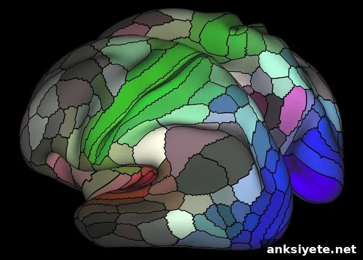
Summary: Researchers have created new software that can accurately identify and map distinct areas of the cerebral cortex.
Source: NIH / NIMH
Software Automatically Identifies Distinct Cortical Areas in Brain Scans
Researchers have produced a detailed map of the human cerebral cortex that identifies 180 distinct areas in each hemisphere — more than twice the number that had been reliably mapped before. Alongside this parcellation, the team developed software that automatically recognizes the unique multi-modal “fingerprint” of each cortical area in an individual’s brain scans. Supported by the National Institutes of Health through the Human Connectome Project (HCP), this approach integrates multiple non-invasive MRI measures that validate one another, producing highly reliable anatomical delineations.
“These new maps and tools provide fresh insight into how the cortex evolved, the specialized roles of its areas in health and disease, and could eventually improve precision in neurosurgery and clinical assessment,” said Bruce Cuthbert, Ph.D., acting director of the NIH’s National Institute of Mental Health (NIMH), which co-funded the research as part of the HCP.
The study identified 97 previously unrecognized cortical areas per hemisphere while confirming 83 areas documented in earlier work, achieving an overall detection rate near 97 percent.
The research team, led by David Van Essen, Ph.D., and Matthew Glasser, Ph.D., at Washington University in St. Louis and collaborators at multiple centers, published their findings in Nature on July 20, 2016. Their multi-modal strategy overcomes limitations of prior studies that relied on single measures — for example, postmortem microscopy alone — which sometimes produced inconsistent borders between cortical areas and reduced comparability across studies.
Glasser likened prior uncertainty in cortical mapping to blurred astronomical images created by ground-based telescopes before adaptive optics and space telescopes improved resolution. To remove that blurriness, the HCP team combined several precisely aligned MRI modalities in a group of 210 healthy young adults. The measures included cortical thickness, myelin content, task-based functional MRI (fMRI), resting-state fMRI, connectivity patterns, and topographic organization. Each measure cross-validated the others, and the resulting parcellation was tested and confirmed in an independent sample of 210 additional participants.
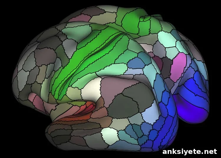
Although a small fraction of participants showed atypical placement of some cortical areas, the data-driven algorithms in the software were still able to detect and correctly map those regions. While the study used task-based fMRI as well as resting-state scans, the researchers concluded that resting-state MRI alone should be sufficient for future mapping with the new tools. The authors also acknowledged that additional data could reveal further subdivisions or hierarchical relationships among some areas.
Glasser emphasized that “the ability to separate individual differences in the location, size, and topology of cortical areas from differences in their activity or connectivity will help clarify how structural and functional features relate to behavior and genetics.”
The team released an automated “areal classifier” and supporting tools to the research community through HCP resources. The extensively analyzed datasets underlying the published figures are available through the NIH-funded database maintained by the Van Essen laboratory, facilitating replication and further studies.
Funding: Supported by the National Institutes of Health, including the NIH / National Institute of Mental Health.
Reporting: Jules Asher, NIH / NIMH
A multi-modal parcellation of human cerebral cortex
Accurate understanding of the human cerebral cortex requires a detailed map of its major subdivisions, referred to as cortical areas. Using multiple aligned MRI modalities from the Human Connectome Project and an objective semi-automated neuroanatomical approach, the authors delineated 180 areas per hemisphere in a precisely aligned group average of 210 healthy young adults. They characterized 97 new areas and confirmed 83 areas previously described through postmortem microscopy and other approaches. To enable automated identification of these areas in new subjects and future studies, a machine-learning classifier was trained to recognize the multi-modal fingerprint of each cortical area. This classifier detected 96.6% of cortical areas in new subjects, replicated the group parcellation, and could correctly locate areas in individuals with atypical parcellations. The freely available parcellation and classifier will improve neuroanatomical precision for studies of cortical structure and function, and for investigations of individual variability, development, aging, and disease.
Paper: “A multi-modal parcellation of human cerebral cortex” by Matthew F. Glasser, Timothy S. Coalson, Emma C. Robinson, Carl D. Hacker, John Harwell, Essa Yacoub, Kamil Ugurbil, Jesper Andersson, Christian F. Beckmann, Mark Jenkinson, Stephen M. Smith, and David C. Van Essen. Published in Nature, July 20, 2016.