
Summary:
Researchers at the Allen Institute have overturned a long-standing idea about how the brainstem nucleus called the locus coeruleus (LC) operates. Rather than acting as a broad, indiscriminate broadcast that floods the brain with norepinephrine, the LC appears to route highly specific norepinephrine signals to distinct anatomical targets—more like a targeted postal network than a foghorn. The team also reconstructed individual LC neurons and identified one axon exceeding 70 centimeters, the longest single neuron recorded in a mouse.
Key Facts:
- Targeted Delivery, Not a Foghorn: Upper (dorsal) LC neurons project primarily to the cerebral cortex and carry signals important for learning, while lower (ventral) LC neurons send projections down to the brainstem and spinal cord and influence basic engagement with the environment.
- Record-Breaking Axon Length: The average LC axon length measured about 35 centimeters; one reconstructed LC neuron had an axon 70.32 cm long—the longest neuronal projection documented in a mouse.
- Therapeutic Implications: Because LC neurons are among the earliest to degenerate in Alzheimer’s disease and because norepinephrine pathways are targets for medications used in ADHD, depression, and anxiety, the detailed circuit map could guide therapies that modulate specific LC subcircuits instead of broadly altering norepinephrine across the entire brain.
Source: Allen Institute
Beyond the Loudspeaker Model
The locus coeruleus is a small, pigmented cluster of norepinephrine-producing neurons deep in the brainstem. Despite its modest size, it exerts outsized influence over attention, arousal, stress responses, heart rate, and learning.
For many years, the dominant view held that LC neurons acted as a uniform broadcast system, releasing a generalized surge of norepinephrine across the brain in response to novelty or stress. The new, multi-method study published in Nature challenges that idea.
Using large-scale whole-brain imaging, electrophysiology, single-cell genetic profiling, and behaviorally demanding tasks in mice, the Allen Institute team demonstrated that LC neurons are organized topographically and functionally. The LC sends distinct norepinephrine signals to specific targets, and those signals correlate with the cells’ positions and gene-expression profiles.
“The findings suggest the brain’s norepinephrine system is far more like a targeted postal network than a foghorn,” said Karel Svoboda, Ph.D., director of Neural Dynamics at the Allen Institute and a co-author of the study.
Segregated Circuits for Learning and Engagement
By tracing axonal paths and examining genetic markers, the researchers identified a clear spatial and functional division within the LC:
- Dorsal LC Neurons: These upper LC cells send projections to the forebrain and isocortex. They become active when animals adjust choices following negative feedback and appear to carry reward-prediction-error signals that drive learning and behavioral adaptation.
- Ventral LC Neurons: These lower LC cells project to the brainstem and spinal cord. They show elevated background activity when animals disengage or ignore cues that indicate available rewards, implicating them in basic states of engagement and environmental responsiveness.
“What emerged was a clear map: neurons in the dorsal LC that send signals upward to the cortex are involved in learning, whereas neurons in the ventral region projecting downward to the brainstem and spinal cord govern whether animals engage with their environment,” Svoboda explained. The team also found accompanying differences in gene expression that align with these anatomical and functional distinctions.
The Longest Axon in the Mouse Brain
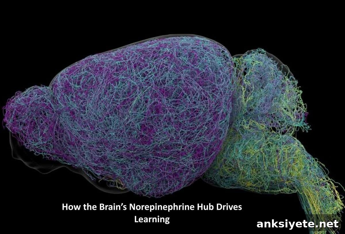
Comprehensive whole-brain imaging of nearly 35,000 neurons, together with genetic profiling of roughly 400,000 cells, allowed full morphological reconstructions of individual LC projections. Those reconstructions revealed exceptionally large projection territories and surprisingly long axons.
On average, LC axons were about 35 centimeters long. One neuron stood out with an axon measuring 70.32 centimeters—over 27 inches—making it the longest individual neuron ever recorded in a mouse. Despite their size, these long-projecting LC neurons do not broadcast uniformly; they selectively innervate cortical regions while sparing structures such as the cerebellum, brainstem, and spinal cord.
“This neuron, like many others that we studied, supplies norepinephrine to a very large volume of the cerebral cortex. For the brain, this is highly unusual. Most neurons are more specific in their targets,” said Jeremiah Cohen, Ph.D., a co-author and scientist at the Allen Institute.
A Dual-Engine Learning Platform
The authors draw parallels between the LC norepinephrine system and the brain’s dopamine pathways. Dopamine relays reinforcement signals primarily to the basal ganglia to shape habits, while dorsal norepinephrine projections deliver error and learning signals to the cortex. Together, these systems form a coordinated computational platform that supports flexible learning and the acquisition of complex, abstract rules.
Clinically, these structural and functional insights matter. The LC degenerates early in Alzheimer’s disease, and many psychiatric medications influence norepinephrine signaling. This detailed map points toward future strategies that could selectively target LC subcircuits for neuroprotective treatments or psychiatric interventions, limiting off-target effects that come from broadly modulating norepinephrine.
Funding: Supported by the National Institutes of Health’s BRAIN Initiative.
Editorial Notes:
- This article was edited by a Neuroscience News editor.
- The original journal article was reviewed in full for accuracy.
- Additional explanatory context was provided by editorial staff.
About this Neuroscience and Learning Research:
- Media Contact: Peter Kim
- Source: Allen Institute
- Image Credit: Image credited to Allen Institute
- Original Research: Nature (September 16, 2026). Title: “Topographic structure and function of locus coeruleus noradrenaline neurons.” Authors include Zhixiao Su, Polina Kosillo, Kanghoon Jung, Shuonan Chen, Mathew T. Summers, Alex Piet, Han Hou, and many colleagues. DOI: 10.1038/s41586-026-11026-0
Abstract
Topographic structure and function of locus coeruleus noradrenaline neurons
Noradrenaline (also known as norepinephrine) is released throughout most of the central nervous system by neurons in the locus coeruleus. In this study, researchers identified relationships among neuronal morphology, gene expression, and activity in mouse LC cells. Individual axonal projections were extensive but largely confined to specific subsets of brain regions. Projection patterns and graded gene expression correlated with the locations of cell bodies within the LC. In a behavioral paradigm requiring continuous learning from actions, dorsal LC neurons projecting to isocortex were activated when mice switched choices and by reward-prediction-error signals that drive learning. Background activity in ventral LC neurons was elevated when mice ignored stimuli indicating potential reward availability. These observations reveal a topographically organized neurotransmitter system that contains learning signals essential for flexible behavior.