
Summary:
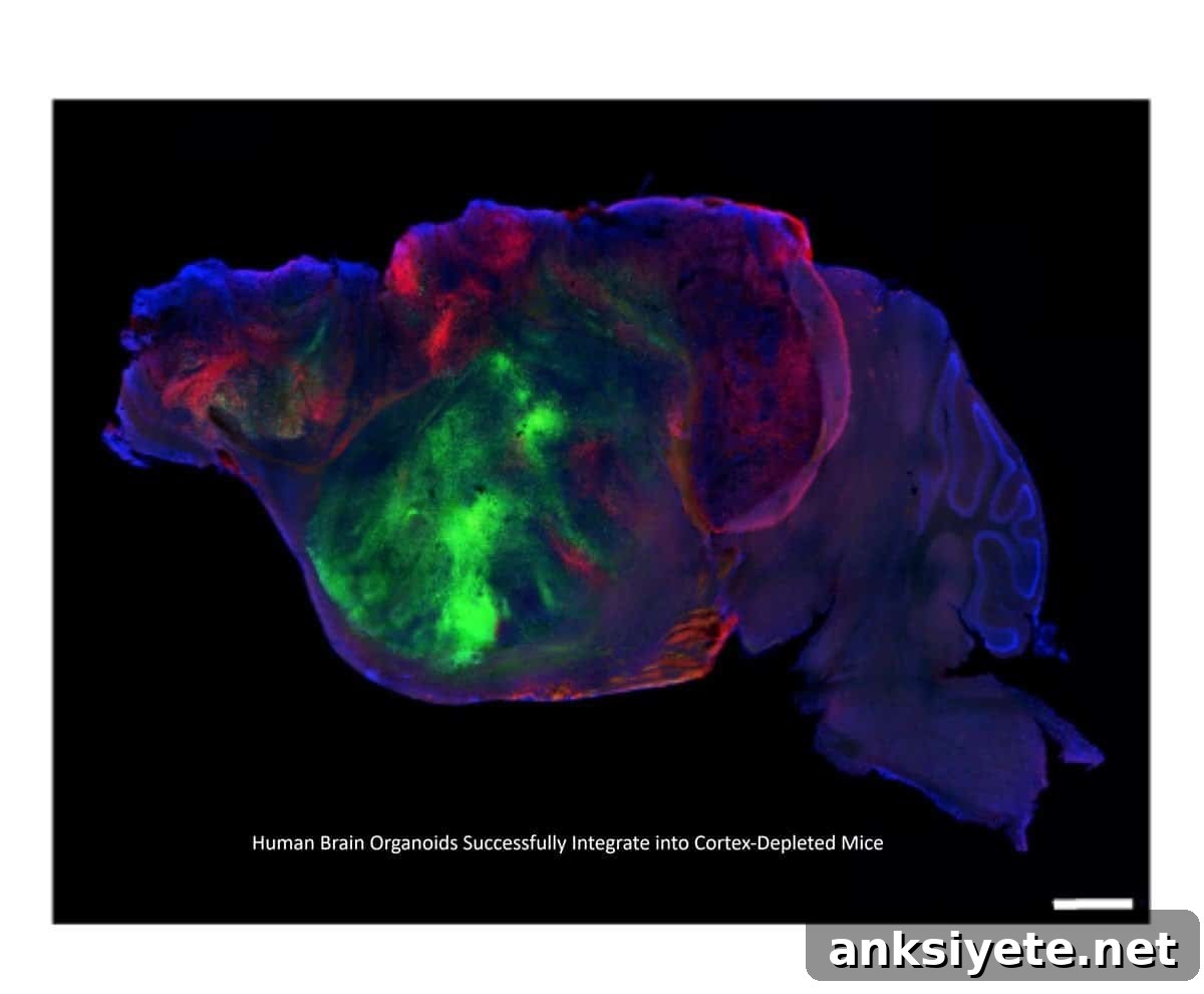
Neuroscientists have created a genetic approach that produces mice missing the majority of their cerebral cortex, opening a large physical niche for transplanted human brain organoids to expand, integrate, and mature. The engrafted human tissue developed into diverse neuronal and glial populations—including cell types that are difficult or impossible to obtain in standard laboratory cultures—and formed functional, circuit-level connections with the host brain and spinal cord that impacted behavior.
Key Facts:
- Cortex Depletion Creates Structural Room: Researchers used genetic engineering to generate mice that develop without most of their cerebral cortex, overcoming cranial space constraints and competition from host neurons so human organoids can occupy and rebuild cortical structures.
- Cellular Diversity and In Vivo Maturation: Transplanted human organoids differentiated into a broad spectrum of cortical cell types, including rare neurons and glia that fail to mature in conventional in vitro systems, and established long-range projections into the mouse central nervous system.
- Behavioral and Disease Modeling Impact: Mice bearing human organoid grafts showed measurable changes in motor coordination and memory tasks compared with cortex-depleted controls, demonstrating a living platform to model injuries and neurodevelopmental disorders.
Source: Stanford University / Nature
Studying human cortical development, psychiatric disorders, and neurodegeneration has been limited by the inaccessibility of living human brain tissue. Three-dimensional stem-cell-derived brain organoids have provided a powerful experimental tool, but when cultivated in dishes they are constrained by lifespan, lack of robust vascularization, and incomplete circuit maturation.
Previous efforts to transplant human organoids into rodents partially addressed these limitations, yet physical bottlenecks persisted: fixed cranial boundaries and dense native neural circuits limited graft expansion and functional integration. In response, a team led by Dr. Sergiu Pașca published a study in Nature describing a developmental engineering strategy that circumvents those constraints. By genetically depriving mice of the majority of their cortical glutamatergic neurons, the researchers created a neonatal cortical cavity that human stem-cell-derived organoids could occupy and develop within.
Cellular Differentiation Beyond the Petri Dish
When human cortical organoids were engrafted into the cortex-depleted space, the grafts expanded to fill the cavity and organized into layered architectures resembling human cortex. The in vivo environment supplied biological signals missing from incubators—vascular support, host-derived trophic factors, and experience-dependent activity patterns—enabling maturation of multiple neuronal and glial subtypes.
Importantly, the human neurons did not remain isolated. They extended axonal projections throughout the host nervous system. Electrophysiology and anatomical tracing showed that graft neurons formed synaptic connections with host circuitry, reaching the brainstem and extending into the spinal cord. These findings demonstrate that human-derived cortical tissue can become functionally embedded in a living mammalian nervous system and participate in circuit-level signaling.
Behavioral Rescue and Disease Modeling
To determine whether the engrafted human tissue produced measurable effects on host behavior, the investigators tested the animals in a series of behavioral paradigms. Mice carrying human organoid grafts exhibited distinct differences in motor coordination and certain memory measures compared with cortex-depleted mice that did not receive grafts. Locomotion overall remained largely intact, but limb coordination and spontaneous behavior organization showed selective alterations tied to the presence of human cortical tissue.
The platform also proved useful for modeling injury to developing human neurons. When researchers induced hypoxic injury—a condition relevant to perinatal stroke and cerebral palsy—the chimeric animals displayed subtle but quantifiable changes in gait and locomotion, indicating that human cellular insults can be translated into organism-level behavioral readouts. This capability supports using xenocortical models to study human neurodevelopment, disease mechanisms, and therapeutic screening at the circuit and behavioral levels.
Navigating the Ethics of In Vivo Brain Models
Because these xenocortical models blur conventional lines between human cellular architecture and animal physiology, the research team emphasized strict adherence to specialized ethical oversight. They recommend continued, proactive dialogue among scientists, bioethicists, regulators, and the public to develop clear policies governing research with human organoid-animal chimeras and to address societal and clinical concerns.
Looking ahead, the authors hope this approach accelerates translational work—allowing drugs and interventions to be tested directly on developing human neural circuits within a living host—while preserving careful ethical governance.
Editorial Notes:
- This article was edited by a Neuroscience News editor.
- Journal paper reviewed in full.
- Additional context added by our staff.
About this Genetics and Neurology Research:
- Media Contact: Sergiu Pașcal
- Source: Nature
- Image Credit: Image credited to S. Pasca lab, Stanford University / Nature
- Original Research is Open Access: Nature (September 16, 2026). “Developmental xenocortication using human-derived organoids in mice.” Authors: Konstantin Kaganovsky, Kevin W. Kelley, Tilo Gschwind, Paul M. Harary, John Kochalka, Alexander D. White, Garikoitz Lerma-Usabiaga, Xiaoyu Chen, Omer Revah, Felicity Gore, Ayano Aoyama, Jennifer L. Shadrach, Se-Jin Yoon, Alfredo Valencia, Satoe Ogawa, Noah Reis, Hannes Vogel, Brian Wandell, Julia A. Kaltschmidt, Ivan Soltesz, Karl Deisseroth & Sergiu P. Pașca.
- DOI: 10.1038/s41586-026-11032-2
Abstract
Developmental xenocortication using human-derived organoids in mice
Limited access to living human brain tissue constrains studies of human development and circuit function; stem-cell-derived organoids are helping bridge that gap. Transplantation of human neural organoids into rodent hosts enables in vivo analysis of human neurodevelopment, circuit dynamics, and host behavioral outcomes.
Spatial constraints and competition with host circuits have previously limited organoid integration and functional study. Here, the authors establish a transplantation platform that uses a genetic method to deplete glutamatergic neurons from mouse neocortex and hippocampus (apallial regions) and to engraft human stem-cell-derived cortical organoids (hCO) neonatally, creating xenocortical mice. Grafts robustly expand to occupy much of the cortical volume and generate diverse human cortical cell types, including layer 5 extratelencephalic projection neurons.
Human cortical neurons integrate with the mouse nervous system. In vivo calcium imaging across cortical grafts and electrophysiological recordings reveal organized activity patterns resembling developing circuits. Behavioural analyses show broadly preserved locomotion with selective differences in limb coordination and altered patterns of spontaneous behaviour. Finally, the platform enables behavioral readouts after injury to developing human cortical cells. Xenocortication offers a route to obtain circuit- and behavior-level readouts using human neurons to study neurodevelopment, model disease, and test therapeutics.