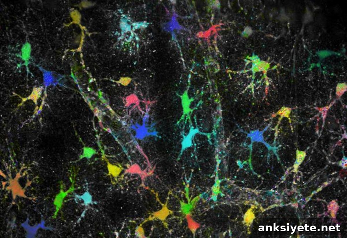
Summary: A new study challenges the long-standing assumption that sodium levels are uniform across astrocytes, the star-shaped glial cells of the brain. Using a newly developed imaging approach, researchers visualized sodium concentrations in real time inside astrocytes and their ultrafine processes for the first time.
The results show that instead of a single static baseline, specialized sodium micro-domains vary dynamically within individual astrocytes and their sub-domains, adjusting to the local excitability demands of neighboring neuronal circuits.
Key Facts
- The glial framework: Glial cells, including astrocytes, make up roughly half of the human brain. They shape brain development, support communication between neurons, and regulate the excitability and proper function of neural networks.
- Electrolyte balance: Sodium ions (Na+), the primary positively charged electrolytes obtained largely from dietary salt, are essential for many physiological processes. In astrocytes, keeping intracellular sodium low is crucial for neurotransmitter regulation at synapses and for managing other ionic balances.
- Revising the uniformity assumption: Neurobiologists previously assumed a consistent, low sodium concentration across all astrocytes and their substructures to preserve reliable housekeeping functions. The team’s direct tissue imaging disproved this view, revealing pronounced variability between cells and within their subregions.
- Membrane transport architecture: Collaborators at Friedrich-Alexander-Universität Erlangen-Nuremberg helped demonstrate that these local sodium differences are driven by membrane transport proteins whose abundance and arrangement vary across astrocyte membranes.
- Multi-scale validation: Experimental observations from isolated brain tissue at HHU were incorporated into biophysical computer models by the University of South Florida and then validated in living animal models by teams at the University of Bonn and University Hospital Bonn.
- Clinical implications: Lead investigators emphasize that these sodium sub-domains respond dynamically to nearby synaptic activity. Disruption of these localized electrolyte balances could contribute to disorders marked by failing ion regulation, including epilepsy and acute stroke, pointing to new therapeutic targets.
Source: HHU
Astrocytes, the star-shaped members of the glial cell family, form a substantial portion of brain tissue and play essential roles in development, synaptic communication, and network stability.
Sodium ions (Na+) are the body’s most important electrolytes and are crucial in many biological processes. Dietary salt (NaCl) is the primary source of these ions. In the brain, sodium concentrations must be tightly controlled because they influence neurotransmitter handling and the balance of other ions that determine neuronal excitability.
Within astrocytes, a low intracellular sodium concentration supports neurotransmitter clearance at synapses and helps regulate extracellular potassium and other electrolytes. These actions enable astrocytes to preserve neuronal function and maintain proper excitability across circuits.
At the Institute of Neurobiology at Heinrich Heine University Düsseldorf (HHU), the team led by Professor Christine R. Rose developed an imaging technique as part of the SynGluCross project (funded by the Federal Ministry of Education and Research) that directly visualizes sodium content in astrocytes and their fine processes in brain tissue. This method permitted quantitative, subcellular observations that were not possible with prior approaches.
Working with colleagues at Friedrich-Alexander-Universität Erlangen-Nuremberg, the University of Bonn, University Hospital Bonn, and the University of South Florida, the researchers tested the prevailing assumption of uniform low sodium across astrocytes. Their experiments revealed marked heterogeneity—both between individual astrocytes and across distinct subdomains within single cells.
Further analysis showed that specific transport proteins in the astrocyte membrane, whose expression patterns and configurations differ between cells and compartments, underlie these localized sodium differences. Biophysical modeling by collaborators in the United States reproduced the experimental patterns, and independent experiments in living animals confirmed the findings observed in isolated tissue.
Dr. Jan Meyer, the study’s lead author, summarized: “We demonstrated specialized functional subdomains within astrocytes created by differing sodium concentrations. Each subdomain adapts to the local requirements of neighboring neural networks.”
Professor Christine R. Rose, the study’s head, added: “These newly identified features of astrocytes likely matter for conditions where ion homeostasis and neurotransmitter regulation break down—such as epilepsy or after a stroke—and they open new avenues for targeted research and potential interventions.”
Key Questions Answered:
A: Astrocytes act as attentive neighbors to neurons. Because synapses in a local network can have varying activity levels, astrocytes establish isolated sodium micro-domains within their fine branches so each region can respond rapidly and specifically to the local demands of nearby neurons.
A: The project used a multi-scale validation strategy: direct imaging of isolated brain tissue in Düsseldorf produced the primary observations; biophysical simulations developed in Tampa reproduced those patterns; and in vivo experiments in Bonn confirmed the localized sodium heterogeneity in living animal models.
A: The findings point to new targets for drug development. Since seizures and stroke involve severe disturbances in ion balances and neurotransmitter dynamics, understanding how astrocytic transport proteins maintain localized sodium domains could enable therapies that stabilize these mechanisms and prevent catastrophic ionic collapse during acute events.
Editorial Notes:
- This article was edited by a Neuroscience News editor.
- The journal paper was reviewed in full by our editorial team.
- Additional context was added by staff for clarity.
About this neuroscience research news
Author: Arne Claussen
Source: HHU
Contact: Arne Claussen – HHU
Image: Image credit: HHU / Institute of Neurobiology – Jan Meyer
Original Research: Open access. Title: Cellular and subcellular heterogeneity of astrocytic Na⁺ homeostasis tuning astrocytes into functionally distinct subgroups in the mouse brain. Journal: Nature Communications. DOI: 10.1038/s41467-026-73435-z
Abstract
Cellular and subcellular heterogeneity of astrocytic Na⁺ homeostasis tuning astrocytes into functionally distinct subgroups in the mouse brain
Astrocytes contribute to extracellular ion and neurotransmitter homeostasis, with the inward Na⁺ gradient playing a central role. Previous work suggested a uniformly low Na⁺ distribution in astrocytes, supporting the notion that these essential homeostatic properties are conserved. Using multiphoton fluorescence lifetime imaging, this study quantitatively measured astrocytic [Na⁺] in mouse brain slices and in vivo.
The data reveal pronounced subcellular and cellular heterogeneity in astrocytic [Na⁺], along with differences in the capacity for Na⁺/K⁺-ATPase (NKA)-mediated uptake of extracellular K⁺. RNAscope and immunohistochemistry indicate differential spatial expression patterns of NKA ß1 and ß2 subunits in astrocytes. Biophysical modeling that incorporates variable NKA expression and differing strengths of Na⁺ influx reproduces the experimentally observed heterogeneity in astrocytic [Na⁺].
Together, these results support the existence of functionally distinct astrocytes and subdomains where Na⁺ homeostasis is locally tuned to meet the specific demands of surrounding neural networks.