
Summary:
Researchers at Mount Sinai have uncovered how the Alzheimer’s genetic risk factor APOE4 actively drives cerebrovascular degeneration. Their work shows APOE4 reprograms vessel-supporting pericytes into scar-forming myofibroblast-like cells, causing vascular fibrosis and promoting amyloid accumulation around blood vessels. At the same time, APOE4 disrupts cholesterol handling in astrocytes, impairing lysosomal waste clearance and enabling toxic protein aggregates to accumulate and spread. Using a novel, cryopreservable 3D human tissue model called “miBrains,” the team demonstrated that blocking TGF-β signaling and targeting cholesterol metabolism can reverse vascular damage and reduce toxic protein buildup in human-relevant tissue models and aged animal models.
Key Facts:
- Vascular fibrosis can be reversed: APOE4 drives a pericyte-to-myofibroblast transition that triggers fibrosis and vascular amyloid deposition. Pharmacological inhibition of the TGF-β pathway restored pericyte coverage and reduced vascular scarring and amyloid in aged APOE4 models.
- Astrocyte cholesterol blocks waste clearance: APOE4 causes abnormal cholesterol buildup in astrocytes, which disrupts lysosomal function and prevents effective clearance of toxic proteins such as alpha-synuclein, allowing them to aggregate and propagate to neurons.
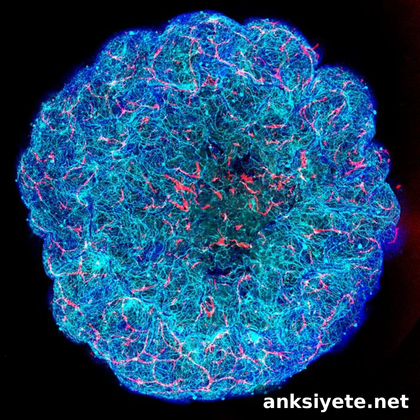
- Scalable, patient-specific miBrains: The team developed a reproducible 3D human tissue platform from induced pluripotent stem cells (iPSCs) that combines neurons, glia, myelinating oligodendrocytes, and functional vasculature to study neurodegenerative mechanisms and test therapeutic strategies in human-like tissue.
Source: The Mount Sinai Hospital / Icahn School of Medicine at Mount Sinai
Alzheimer’s disease affects more than seven million older adults in the United States and progressively erodes memory, cognition, and daily function. Although vascular decline in the brain has long been associated with Alzheimer’s, especially among APOE4 carriers, it has often been treated as secondary damage. New studies from Mount Sinai overturn this view, showing cerebrovascular deterioration is an active, APOE4-driven process that can be intercepted.
Published as complementary papers in Cell and Cell Stem Cell, the new research identifies cellular and molecular pathways by which APOE4 undermines the neurovascular unit and worsens proteinopathy. These discoveries point to actionable targets—TGF-β signaling in pericytes and cholesterol handling in astrocytes—that could preserve vascular health and limit toxic protein spread in Alzheimer’s and related disorders.
“Damage to the brain’s blood vessels is not simply a late consequence of Alzheimer’s; it is a biologically active process caused by APOE4 that may be reversible,” says corresponding author Joel W. Blanchard, PhD, Associate Professor of Neuroscience and Stem Cell Biology and Regenerative Medicine at the Icahn School of Medicine at Mount Sinai. “These findings reveal therapeutic strategies to preserve cerebrovascular function and reduce amyloid buildup in people at genetic risk for Alzheimer’s disease.”
Transforming Vessel Support Cells into Scar Tissue
In the Cell paper, Mount Sinai scientists created a single-cell transcriptomic atlas of the human brain vasculature to map gene expression across cells that build and maintain the blood-brain barrier. Their analysis showed that APOE4 is linked to a marked reduction in normal pericytes and the appearance of a myofibroblast-like population expressing contraction and extracellular matrix genes.
Pericytes normally wrap capillaries and preserve vessel integrity. Under the influence of APOE4, those pericytes adopt a scar-forming identity, secreting fibronectin and other extracellular matrix proteins that stiffen and fibrose vessels. That fibrosis promotes the accumulation of vascular amyloid-beta, further compromising circulation and tissue health.
Using immunostaining in human and mouse brain samples, the team validated the presence of non-vascular myofibroblasts associated with APOE4. Computational and experimental work implicated elevated transforming growth factor β (TGF-β) signaling as the key driver of the pericyte-to-myofibroblast transition. Importantly, inhibiting TGF-β signaling in aged APOE4 models restored pericyte coverage, reduced vascular fibrosis, and lowered perivascular amyloid to levels more typical of APOE3, demonstrating that this maladaptive vascular remodeling is druggable and reversible.
Astrocyte Cholesterol and Cellular Waste Failure
In the companion Cell Stem Cell study, investigators used the miBrains platform—3D human tissue constructs derived from iPSCs that include neurons, oligodendrocytes, astrocytes, and functional vasculature—to examine how APOE4 fosters toxic protein aggregation. The model allowed real-time study of human cellular interactions driving neurodegeneration.
The researchers identified a cascade of metabolic disruption in APOE4 miBrains:
- Cholesterol congestion: APOE4 causes abnormal cholesterol accumulation inside astrocytes, altering lipid distribution across the neurovascular unit.
- Lysosomal impairment: Excess cholesterol impairs astrocyte lysosomes, crippling their ability to degrade and clear misfolded or aggregated proteins.
- Pathological propagation: With glial clearance disabled, toxic alpha-synuclein accumulates, aggregates, and spreads into nearby neurons, seeding neurodegenerative inclusions.
These results nominate astrocyte cholesterol trafficking and lysosomal degradation as therapeutic targets to prevent spread of toxic protein aggregates across Alzheimer’s disease, Parkinson’s disease, and Lewy body dementia.
Scalable, Patient-Specific “miBrains”
A central challenge for neurodegenerative research is modeling living human brain pathology before irreversible postmortem changes occur. miBrains address that gap by recreating human neurovascular pathology in a scalable and reproducible format. The platform can be generated with defined cell compositions and disease-associated factors, then cryopreserved for consistent use in experiments and drug screens.
“A key advance of our technology is that miBrains with predefined cellular compositions and disease-related features can be cryopreserved,” says Louise Mesentier-Louro, PhD, Assistant Professor of Neuroscience and Stem Cell Biology and Regenerative Medicine and first author on the Cell Stem Cell paper. “This improves reproducibility and throughput for complex disease modeling and accelerates preclinical testing of therapeutic candidates.”
Mount Sinai is producing patient-derived miBrains to build a personalized testing platform for assessing disease vulnerability and drug efficacy, creating a direct bridge from genetic discovery to individualized therapeutic evaluation.
Funding: The Cell study on cerebrovascular degeneration received support from NASA, the National Institute on Aging (NIH), The SWT Foundation, and the CureAlz Fund. The Cell Stem Cell study on abnormal protein accumulation was supported by NASA, Aligning Science Across Parkinson’s (ASAP), the Michael J. Fox Foundation, the National Institute of Neurological Disorders and Stroke, the National Institute on Aging (NIH), The SWT Foundation, and the CureAlz Fund.
Editorial Notes:
- This article was edited by a Neuroscience News editor.
- Journal paper reviewed in full.
- Additional context added by staff.
About this Depression Research:
- Media Contact: Stacy Anderson
- Source: Mount Sinai Hospital
- Image Credit: Image credited to The Blanchard Lab / The Mount Sinai Health System
- Original Research is Open Access: Cell (September 24, 2026). “A pericyte-to-myofibroblast transition links APOE4 to cerebrovascular degeneration.” Authors: Braxton R. Schuldt et al.
- DOI: 10.1016/j.cell.2026.08.058
Abstract
A pericyte-to-myofibroblast transition links APOE4 to cerebrovascular degeneration
Cerebrovascular disease is a prominent but understudied feature of Alzheimer’s disease (AD). The strongest genetic risk factor for AD, apolipoprotein E4 (APOE4), is associated with vascular amyloid deposition and fibrosis. To define how APOE4 drives cerebrovascular pathology, the investigators assembled a single-cell transcriptomic atlas of the human brain vasculature. They found that APOE4 carriers show reduced pericyte abundance and the emergence of a myofibroblast-like population that co-expresses contractile and extracellular matrix genes.
Immunostaining validated the presence of non-vascular myofibroblasts in APOE4 human and mouse brains. Experimental evidence demonstrated that APOE4 pericytes transition into myofibroblasts that secrete fibronectin, promoting vascular amyloid accumulation. Computational and laboratory analyses identified elevated transforming growth factor β (TGF-β) signaling as the driver of this transition. Pharmacological inhibition of TGF-β restored pericyte coverage and reduced vascular fibrosis and amyloid to levels comparable to APOE3, revealing a targetable mechanism that connects APOE4 to cerebrovascular pathology in Alzheimer’s disease.