
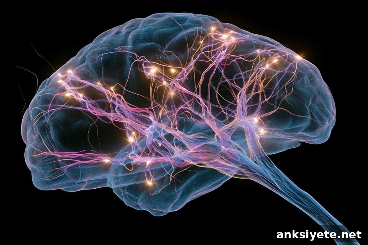
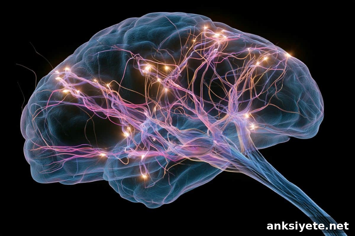
Summary: Researchers have identified an evolutionarily ancient circuit in the brainstem that functions as a core “attentional selection engine.” Long-held views placed selective spatial attention primarily in the prefrontal cortex, a region prominent in primates and humans. That model did not explain how birds, fish, and reptiles successfully filter distractions. New experiments in mice reveal a conserved brainstem mechanism that controls distractibility and target selection, with important implications for understanding attention disorders in humans.
Using a human-like visual attention task, the team at Johns Hopkins University found that a defined population of inhibitory brainstem neurons governs whether an animal prioritizes a central, goal-relevant stimulus over competing peripheral inputs. When researchers temporarily silenced these neurons, mice became markedly distractible: even faint peripheral flashes drew them away from the target. Restoring activity in the same neurons reversed the effect and restored the mice’s ability to ignore strong distractors. Crucially, these changes occurred without impairing vision or motor control, demonstrating that the circuit specifically mediates competitive information processing rather than sensory or motor function.
Key Facts
- The Brainstem Engine: An inhibitory neuronal circuit in the brainstem actively evaluates competing sensory inputs and selects the stimulus that should be attended to immediately.
- Evolutionary Conservation: This attentional mechanism predates the prefrontal cortex by hundreds of millions of years, explaining how species without a large cortex can still focus attention effectively.
- Reversible Hyper-Distractibility: Temporarily deactivating these neurons produced a rapid, reversible state of hyper-distractibility in mice, a behavioral change that closely resembles a core symptom of ADHD.
- Pure Attentional Deficit: Control experiments demonstrated that the deficit from circuit silencing was specific to decision-making among competing stimuli, not due to visual loss or motor impairment.
- Clinical Implications for ADHD and Autism: Since these brainstem neurons are conserved across vertebrates, functional disruptions in the same circuit in humans may contribute to attention-related disorders, suggesting potential for targeted, non-stimulant therapies.
Source: JHU
Neurons tucked inside an ancient brain region suppress distractions and guide focus
Johns Hopkins researchers discovered this population of brainstem neurons in mice, in a region that exists throughout vertebrates, including humans. The finding offers a potential path toward more specific therapeutic strategies for attention disorders.
“A hallmark of ADHD is that even faint distractors draw attention away—and that’s exactly what we see when these neurons are silenced,” said senior author Shreesh Mysore, a neuroscientist who studies neural circuits underlying behavior. “When the neurons are turned back on the next day, the same animal can once again ignore distractors, even strong ones.”
The federally funded study appears in Nature Communications and has been selected as an editorial highlight. Selective spatial attention allows animals and people to focus on relevant information—finding a friend in a crowd or following a conversation in noise—and is impaired in conditions like autism and Attention-Deficit/Hyperactivity Disorder (ADHD).
Traditionally, attention has been attributed mainly to the prefrontal cortex, a brain area especially developed in primates. That explanation leaves unanswered how species without a large prefrontal cortex, such as birds or fish, can nevertheless display focused behavior. Lead author Ninad Kothari, a postdoctoral fellow in the Department of Psychological and Brain Sciences, explained that the team traced this capability to an evolutionarily old brainstem region that supports spatial selection across vertebrates.
In the experimental task, mice were trained to focus on visual information shown centrally while ignoring distracting flashes on the side. Successful responses required touching a location indicated by the central cue rather than the distractor. Mice performed well under normal conditions but became highly distractible when the identified brainstem neurons were temporarily silenced.
The authors ran additional controls to confirm that impairment was not due to reduced vision or motor function. “The only deficit was in comparing competing pieces of information and selecting the location with the most relevant signal,” Mysore said. “This area of the brain acts like an attentional selection engine, answering the question: ‘What is the most important information to focus on right now?’”
Next, the researchers plan to probe how these brainstem neurons exert control over spatial attention across vertebrates and to what extent they contribute to human attention. All current evidence suggests these neurons exist in humans, but whether they play the same central role in selective spatial attention remains an open and testable hypothesis.
If human studies show similar functional involvement, measuring activity in this brainstem circuit in people with ADHD or autism could identify specific dysfunctions and guide development of precision treatments that act locally on this ancient attentional system.
Authors on the paper include Arunima Banerjee, Qingcheng (Jessica) Zhang and Wen-Kai You, together with Kothari and Mysore, all affiliated with Johns Hopkins University.
Key Questions Answered:
A: For decades scientists emphasized the prefrontal cortex as the central seat of complex attention. Identifying a core inhibitory circuit in the ancient brainstem shows that basic spatial attention is an evolutionarily conserved function. This finding explains how animals without a large cortex can perform winner-take-all selection to focus on prey or avoid predators.
A: When the brainstem circuit was disabled, mice became acutely hyper-distractable. During a visual attention task they normally performed reliably, the animals lost focus as soon as minor peripheral distractors appeared. The effect was fully reversible: reactivating the neurons restored normal attention the following day.
A: Current treatments for attention deficits often target widespread neurotransmitter systems in the cortex and can cause off-target side effects. If the same brainstem circuit is implicated in human attention disorders, it could enable development of localized, precision therapeutics that directly modulate an evolutionarily fundamental attentional mechanism.
Editorial Notes:
- This article was edited by a Neuroscience News editor.
- The journal paper was reviewed in full.
- Additional context was added by staff for clarity.
About this neuroscience and ADHD research news
Author: Jill Rosen
Source: JHU
Contact: Jill Rosen – JHU
Image: Image credited to Neuroscience News
Original Research: Open access. “Evolutionarily old brainstem neurons are required for the control of selective spatial attention” by Ninad B. Kothari, Arunima Banerjee, Qingcheng Zhang, Wen-Kai You & Shreesh P. Mysore. Nature Communications. DOI: 10.1038/s41467-026-72340-9
Abstract
Evolutionarily old brainstem neurons are required for the control of selective spatial attention
To behave adaptively in complex environments, animals must selectively process the most important information in space while ignoring distractors.
Here, we report that an evolutionarily ancient group of brainstem inhibitory neurons, called PLTi, is critical for selective spatial attention. In freely behaving mice performing a human-like spatial attention task, bilateral silencing of PLTi severely disrupted target selection without causing perceptual or task-relevant motor impairments.
PLTi’s effects depended on goal-relevant signals rather than mere physical salience, revealing it as a specialized site for priority-driven attentional selection. PLTi primarily controlled the accuracy and categorical precision of the decision boundary that separates the target from lower-priority distractors.
PLTi influenced neural representations of competing stimuli in the superior colliculus, an established attentional hub, suggesting a mechanistic pathway. PLTi may therefore be a conserved brainstem locus across vertebrates for winner-take-all spatial decisions.