
New method from Case Western Reserve and Cleveland Clinic improves prediction of surgical success for temporal lobe epilepsy
Researchers at Case Western Reserve University and Cleveland Clinic have developed a more accurate way to identify the parts of the brain affected by temporal lobe epilepsy, offering patients and clinicians clearer guidance on whether surgery is likely to help. Their findings, published in PLOS ONE, show that measuring functional connectivity with intracranial EEG (iEEG) can predict surgical outcome with substantially higher accuracy than current techniques.
Assistant Professor Roberto Fernández Galán, PhD, led the study that demonstrates how analysis of neuronal activity in the temporal lobe reveals which regions are diseased and therefore good candidates for resection. “Our analysis of neuronal activity in the temporal lobe allows us to determine whether it is diseased, and therefore, whether removing it with surgery will be beneficial for the patient,” Galán said. He described the new approach as a meaningful improvement in both accuracy and efficiency compared to existing methods.
Temporal lobe epilepsy is one of the most common forms of focal epilepsy. For about one-third of people with this condition, medications fail to control seizures. Surgical removal of part or all of the temporal lobe (lobectomy) is a standard treatment option, but success rates have varied, typically reported in the 60–70 percent range. One major challenge is accurately identifying the epileptogenic tissue before surgery so that resection removes the problematic region while preserving healthy function.
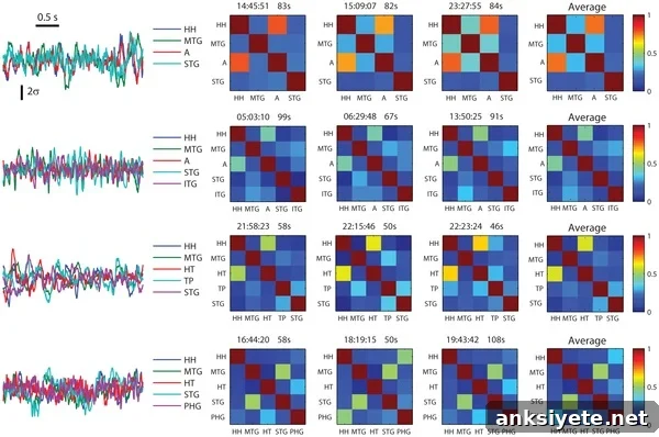
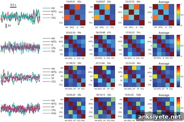
In this retrospective study, investigators analyzed preoperative intracranial EEG data from 23 patients who underwent temporal lobe resections after iEEG monitoring at Cleveland Clinic. Instead of focusing solely on overt epileptic spikes and seizure events, the team quantified functional connectivity—the pattern of communication among different brain regions—during interictal periods (times without visible epileptic discharges). They discovered that characteristics of neural networks, specifically the presence of relatively weak and homogeneous connections within a lobe, reliably indicated diseased tissue.
The research team reports that functional connectivity estimated from iEEG identified the epileptic lobe with 87 percent accuracy in their sample. Arun Antony, MD, the paper’s first author, noted that although functional connectivity has been extensively investigated in basic neuroscience, it has not yet been widely integrated into routine clinical practice for epilepsy. “Our discovery is another step towards the use of measures of functional connectivity in making clinical decisions in the treatment of epilepsy,” Antony said.
This approach differs from conventional preoperative assessment methods that emphasize spikes and synchronous discharges visible in iEEG traces. By extracting meaningful information from background neural activity, the connectivity-based analysis offers an additional objective biomarker that could complement existing diagnostic tests. That has potential value for surgical planning: better identification of the epileptogenic lobe can increase the likelihood that resection will improve seizure control while reducing unnecessary removal of healthy tissue.
The multidisciplinary team included investigators from Cleveland Clinic who contributed clinical expertise, patient data, and surgical follow-up information. Co-authors on the study are Arun R. Antony, Andreas V. Alexopoulos, Jorge A. González-Martínez, John C. Mosher, Lara Jehi, Richard C. Burgess, Norman K. So, and Roberto F. Galán. Dr. Galán is a scholar of The Mt. Sinai Health Care Foundation and a former fellow of The Alfred P. Sloan Foundation.
Clinical adoption of functional connectivity measures will require further validation in larger, prospective cohorts and integration into preoperative workflows. Nonetheless, these results highlight the promise of combining advanced signal analysis with intracranial recordings to personalize surgical decision-making for patients with intractable temporal lobe epilepsy.
Contact: Jessica Studeny – Case Western Reserve University
Source: Case Western Reserve University press release
Image credit: Arun R. Antony, Andreas V. Alexopoulos, Jorge A. González-Martínez, John C. Mosher, Lara Jehi, Richard C. Burgess, Norman K. So, and Roberto F. Galán / PLOS ONE
Original research: “Functional Connectivity Estimated from Intracranial EEG Predicts Surgical Outcome in Intractable Temporal Lobe Epilepsy” by Arun R. Antony et al., PLOS ONE. Published online October 30, 2013.