
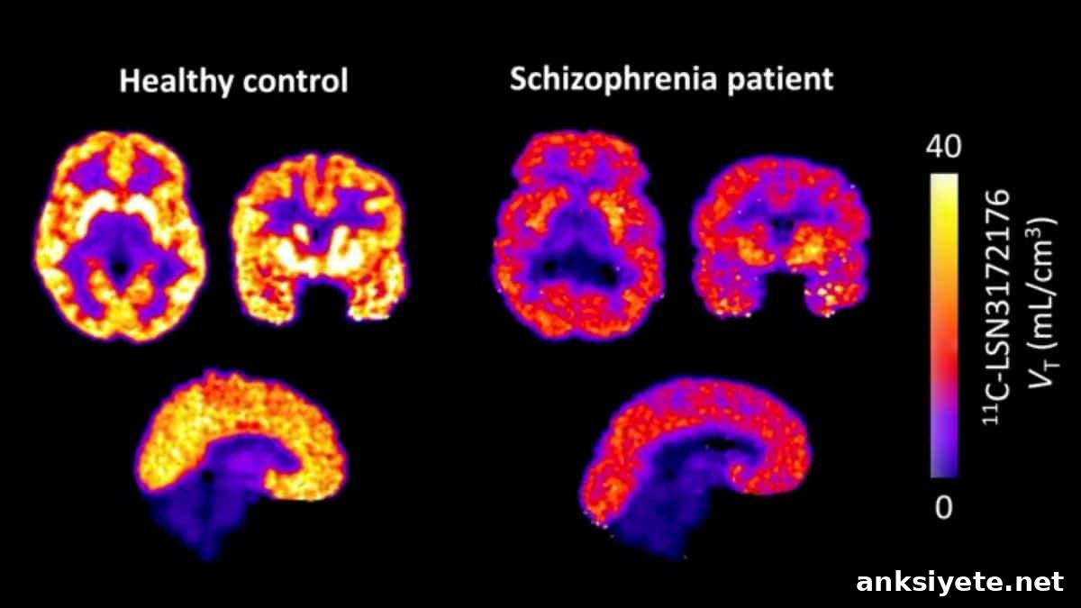
Summary: Researchers have provided the first in vivo evidence of widespread deficits in muscarinic acetylcholine M1 receptors in people with schizophrenia. Using a novel, highly selective PET radiotracer, the team found a marked 13%–19% reduction in M1 receptor availability across multiple brain regions involved in cognition. These findings support a shift toward non-dopaminergic, precision treatments for schizophrenia.
Key Facts
- M1 receptor reductions: High-resolution PET imaging showed that patients with schizophrenia have a consistent 13% to 19% reduction in functional M1 receptor availability across broad cortical and subcortical regions compared with matched healthy controls.
- Connection to cognitive impairment: Lower M1 availability correlated more strongly with objective measures of cognitive dysfunction than with the severity of positive psychotic symptoms, indicating that M1 loss may underlie memory, learning, and executive function deficits.
- First true in vivo confirmation: Prior evidence largely relied on postmortem tissue, leaving open whether deficits existed during life or reflected medication or illness progression. This PET study provides direct in vivo confirmation of M1 deficits in living patients.
- Relevance to non-dopaminergic treatments: The results provide biological support for newly developed muscarinic-based therapies, such as xanomeline–trospium (COBENFY™), which act through muscarinic mechanisms rather than by blocking dopamine D2 receptors.
- Potential for precision psychiatry: Although this study did not measure treatment response, investigators suggest M1 PET imaging could eventually help identify biological subgroups of patients and guide targeted therapies.
- M1 as a drug development target: M1 receptors are widely distributed G-protein-coupled receptors important for synaptic plasticity; these findings strengthen their status as a key therapeutic and diagnostic focus in schizophrenia research.
Source: Elsevier
A new positron emission tomography (PET) study reports significantly lower muscarinic M1 receptor availability—about 13% to 19% lower—in patients with schizophrenia versus healthy individuals. These reductions were observed in brain regions essential for cognition, learning, memory, and executive function.
Published in Biological Psychiatry, the study delivers the first direct in vivo evidence of widespread M1 receptor deficits in schizophrenia.
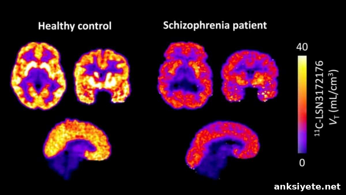
Schizophrenia is a heterogeneous disorder with diverse clinical features and underlying biology. For decades, postmortem studies suggested abnormalities in the brain’s muscarinic acetylcholine system—especially the M1 receptor—but it was unclear whether these deficits existed during life or were secondary to long-term medication and illness effects. The development of a selective PET ligand enabled direct measurement of M1 availability in living patients.
“Developing a selective PET radiotracer for the M1 receptor allowed us to measure receptor availability in the living brain,” explains co-lead investigator Deepak C. D’Souza, MBBS, MD, Yale University School of Medicine. “While availability is not identical to receptor density, it is a recognized proxy for the functional state of the M1 system and allowed us to confirm muscarinic dysfunction in vivo.”
Co-first author Tommaso Volpi, MD, PhD, notes that the results were robust across multiple PET quantification methods and remained significant after accounting for gray matter differences and partial-volume effects.
Investigators emphasize that reduced M1 availability was more closely linked to cognitive deficits than to positive psychotic symptoms. This suggests M1 dysfunction may specifically contribute to the cognitive impairments that often cause the greatest functional disability in schizophrenia.
Traditional antipsychotic treatments target dopamine D2 receptors and are less effective for negative and cognitive symptoms, often producing significant side effects. M1 receptors are G-protein-coupled receptors distributed throughout cortical and subcortical regions and are now receiving attention as promising targets for new therapies and diagnostics.
John Krystal, MD, Editor of Biological Psychiatry, comments that the study is timely given the emergence of muscarinic agonists as schizophrenia pharmacotherapies and the recent approval of xanomeline–trospium (COBENFY™), the first primarily non-dopaminergic antipsychotic approved in decades.
“Although this study did not assess treatment outcomes, it strengthens the biological rationale for muscarinic-based therapies and suggests that M1 receptor imaging could help identify biologically distinct patient subgroups to inform precision medicine approaches,” says co-lead investigator Rajiv Radhakrishnan, MBBS, MD, Yale University School of Medicine.
Key Questions Answered:
A: The limiting factor was not lack of interest but of technology. Visualizing a specific receptor with PET requires a radiotracer that crosses the blood-brain barrier, binds selectively to a single receptor type, and produces a detectable signal. For many years, radiotracers existed mainly for dopamine and serotonin systems; muscarinic receptors were difficult to isolate. The new highly selective M1 radiotracer developed by the research team enabled precise in vivo imaging of the M1 system.
A: Receptor density is the physical count of receptors found on cell surfaces and is typically measurable only in postmortem tissue. Receptor availability, measured by PET, reflects how many receptors are accessible and functional in the living brain. Although not identical, availability is a reliable proxy for the real-time functional state of M1 receptors.
A: For decades, approved antipsychotics have acted primarily by blocking dopamine D2 receptors, which helps positive symptoms but does little for cognitive impairments and often causes notable side effects. Xanomeline–trospium (COBENFY™) works through muscarinic activation rather than dopamine blockade. Showing that living patients have a measurable M1 receptor deficit provides a biological basis for this non-dopaminergic treatment approach.
Editorial Notes:
- This article was edited by a Neuroscience News editor.
- The journal paper was reviewed in full.
- Additional context was added by staff.
About this schizophrenia research news
Author: Eileen Leahy
Source: Elsevier
Contact: Eileen Leahy – Elsevier
Image: Image credited to Biological Psychiatry / Volpi et al.
Original Research: Open access. “Lower Muscarinic M1 Receptor Availability in Schizophrenia: In Vivo PET Evidence” by David Labaree, Deepak C. D’Souza, Mika Naganawa, Nabeel Nabulsi, Rachel Hird, Rajiv Radhakrishnan, Richard E. Carson, Soheila Najafzadeh, Swanee Jacutin-Porte, Tommaso Volpi, Yiyun Huang.
DOI: 10.1016/j.biopsych.2026.06.002
Abstract
Lower Muscarinic M1 Receptor Availability in Schizophrenia: In Vivo PET Evidence
Background
Interest is growing in the role of muscarinic M1 receptors in schizophrenia. Postmortem studies have long suggested M1 deficits, and the recent FDA approval of xanomeline–trospium (an M1/M4 agonist combination) highlights the clinical importance of this system. The PET ligand 11C-LSN3172176 makes it possible to quantify M1 availability in vivo and examine its relationship to clinical features.
Methods
This study compared M1 availability in 16 patients with schizophrenia and 16 age- and sex-matched healthy controls using 11C-LSN3172176 and a high-resolution PET scanner. Measures included distribution volume ratio relative to the centrum semiovale (DVRCS) and distribution volume (VT). Regional gray matter fraction (%GM) was included as a covariate to account for atrophy.
Results
Compared with controls, patients showed significantly lower M1 availability across multiple brain regions with substantial effect sizes: frontal (DVRCS: -13%), temporal (DVRCS: -15%; VT: -12%), parietal (DVRCS: -14%; VT: -11%), occipital (DVRCS: -16%; VT: -13%), caudate (DVRCS: -19%; VT: -15%), putamen (DVRCS: -19%; VT: -17%), hippocampus (DVRCS: -13%), and amygdala (DVRCS: -19%; VT: -16%). A subgroup of patients exhibited larger whole-brain deficits (>20% below control mean). Exploratory analyses indicated associations between M1 availability and selected clinical measures.
Conclusions
These in vivo findings, together with postmortem data, support further investigation of M1 deficits in schizophrenia. The results highlight M1 as both a therapeutic target and a potential biomarker to guide muscarinic-based treatment strategies and precision psychiatry approaches.