
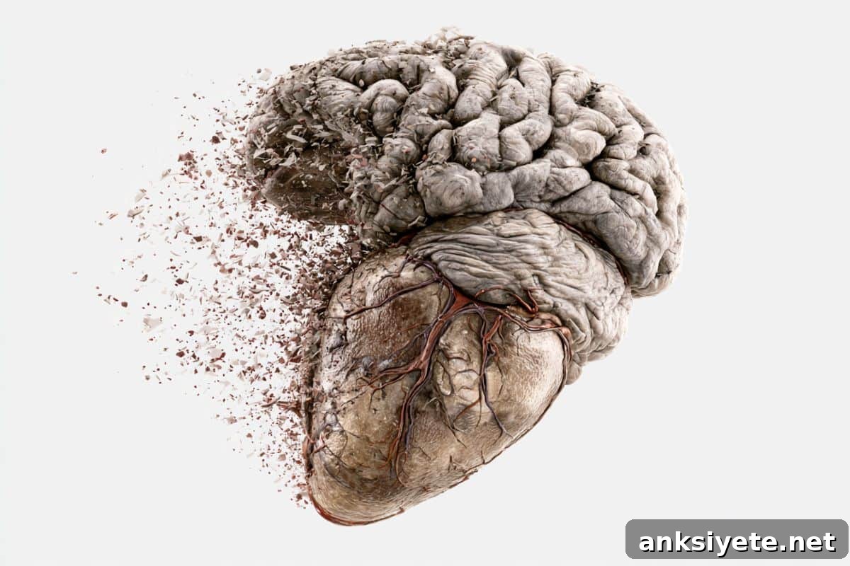
Summary: A longitudinal analysis of 73 participants in the Leipzig Heart Study over 3.5 years found that subtle cardiac dysfunction reliably predicts microstructural tissue loss in brain regions closely linked to Alzheimer’s disease.
Researchers led by Xia Zhang at the Max Planck Institute for Human Cognitive and Brain Sciences report that small reductions in cardiac pumping efficiency — even when not severe enough to qualify as clinical heart failure — were associated with progressive microstructural deterioration of gray and white matter. Those microscopic changes concentrated in memory-related networks and appear to explain why people with subclinical heart problems show measurable long-term memory decline.
Key Facts
- The heart-brain axis: The study highlights an underappreciated connection between cardiac function and brain health. Even minor reductions in cardiac output can produce subtle perfusion deficits that, over time, deprive metabolically demanding brain regions of the oxygen and nutrients they need.
- Alzheimer’s-vulnerable regions: Tissue changes were not widespread or random. Instead, microstructural erosion clustered within the structural networks and temporal lobe regions that are known to be among the earliest targets of Alzheimer’s pathology, particularly structures involved in memory consolidation.
- Microstructure as the missing link: Conventional clinical neuroimaging methods typically measure macro-level features such as total brain volume, major infarcts, or gross atrophy. Those approaches miss early, microscopic tissue changes. By using advanced measures of microstructural integrity, the team identified cellular-level tissue loss that directly mediates the relationship between mild cardiac dysfunction and poorer long-term memory performance.
- Clinical risk stratification opportunity: Zhang and colleagues propose that assessing brain microstructure provides a new, clinically relevant marker for neurologic risk in patients with cardiac impairment. Such metrics could help cardiologists and neurologists identify which patients are most likely to develop cognitive decline and prioritize early intervention.
- Next research steps: Building on the 3.5-year longitudinal data, the investigators plan to incorporate molecular and neural biomarkers into future trials. Their goal is to map the precise overlap between heart-driven brain changes and the earliest, preclinical biological mechanisms that lead to Alzheimer’s disease and related dementias.
Source: SfN
New in Journal of Neuroscience: Xia Zhang and the Max Planck team conducted this longitudinal investigation to clarify how cardiac function and cognition are connected at the tissue level. They followed 73 patients from the Leipzig Heart Study for three and a half years, combining sensitive cardiac assessments with advanced brain microstructure imaging to detect subtle neurodegenerative change.
The primary finding is that subtle cardiac dysfunction — measured below the threshold of clinical heart failure — predicts microscopic deterioration within brain circuits crucial for memory. Those microscopic changes, rather than obvious macrostructural damage, accounted for the observed decline in long-term memory performance among affected participants.
“Monitoring brain microstructural integrity offers a novel and potentially practical approach for neurological risk stratification among patients with cardiac dysfunction,” Zhang said. The implication is clear: routine cardiology evaluations that also consider brain microstructure could enable earlier identification of individuals at risk for cognitive decline.
Current standard imaging — typical hospital MRI or CT scans — is optimized to detect large-scale abnormalities such as tumors, hemorrhages, or advanced atrophy, but it lacks the sensitivity to reveal early cellular-level tissue loss. The methods used by Zhang’s group, by contrast, can detect microstructural alterations before they become apparent on conventional scans, creating an opportunity for earlier, targeted prevention efforts.
Key Questions Answered
A: This study challenges the common assumption that minor cardiac problems affect only physical stamina. The brain is metabolically demanding, consuming a large share of the body’s energy. Even a small, chronic reduction in cardiac perfusion can slowly deprive the brain’s most energy-dependent regions, causing microscopic tissue breakdown in the neural networks that support long-term memory — long before a clinical diagnosis of heart failure arises.
A: Typical clinical scans detect macro-level abnormalities; they are not designed to reveal subtle, cellular-scale changes. The Max Planck team applied advanced imaging techniques focused on microstructural integrity, enabling them to observe early tissue deterioration that standard MRI or CT would miss. Those microstructural changes help explain the cognitive decline observed in patients with mild cardiac impairment.
A: With a clear longitudinal link established, researchers will expand their methods to include specific neural and molecular biomarkers. Future work aims to construct a comprehensive biological map showing how heart-related brain changes intersect with, accelerate, or otherwise influence the molecular cascades that cause Alzheimer’s disease and other dementias. Ultimately, this could inform preventive strategies and targeted therapies for at-risk patients.
Editorial Notes
- This article was edited by a Neuroscience News editor.
- The cited Journal of Neuroscience paper was reviewed in full.
- Additional context was provided by the editorial staff.
About this Alzheimer’s disease research news
Author: SfN Media
Source: SfN
Contact: SfN Media – SfN
Image: The image is credited to Neuroscience News
Original Research: Findings published in Journal of Neuroscience.