
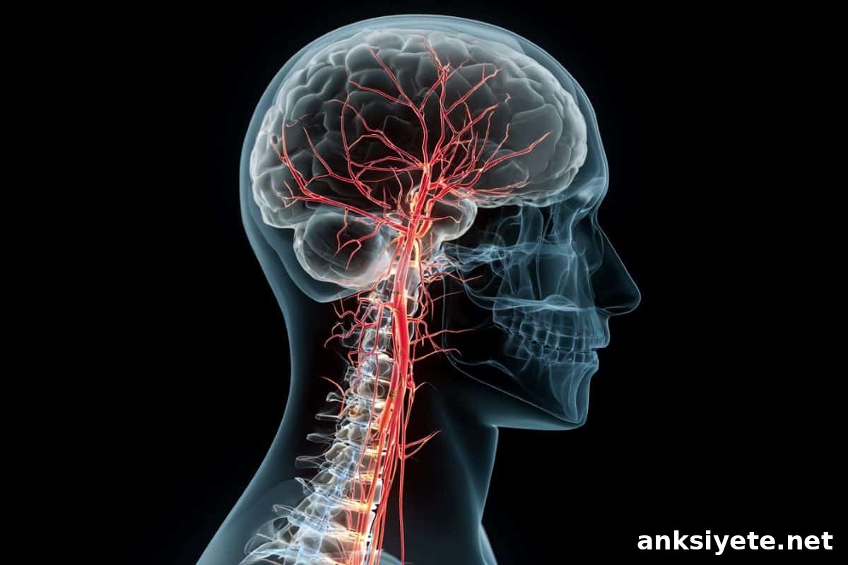
Summary: Many people with incomplete spinal cord injury (SCI) regain the ability to walk, yet everyday tasks such as standing still, balancing, or applying steady force remain challenging. A new study explains how SCI alters the nervous system’s coordination of motor units, producing both shaky low-effort control and overly rigid high-effort responses.
Using non-invasive skin sensors, researchers identified fundamental changes in how motor units—individual nerve-to-muscle elements—are coordinated after incomplete SCI. The team found that at low effort levels the central nervous system fails to synchronize motor units smoothly, which causes instability. At higher effort levels the nervous system appears to overcompensate by sending broad, high-amplitude signals that reduce fine control.
Key Facts
- Coordination breakdown: In people without injury, motor units across muscles receive a common, coordinated neural drive that allows smooth, flexible control. Following incomplete SCI, that shared signal is disrupted.
- Shaky low-effort control: At about 20% of maximal effort, individuals with SCI showed reduced coordination between calf muscles, producing unstable, shaky force output compared with controls.
- Overly loud high-effort signals: At roughly 50% effort, the nervous system in SCI participants matched muscles with stronger low-frequency synchronization, producing a rigid, less precise response.
- Loss of strategic flexibility: Healthy nervous systems adjust the pattern and strength of shared neural drive as force requirements change. After SCI this adaptive flexibility appears reduced, causing a more fixed control strategy.
- Potential rehabilitation biomarker: The altered patterns of neural drive and motor unit synergies may serve as objective biomarkers to guide therapy and to design interventions that re-tune spinal output.
Source: KTH
Even when walking returns, fine motor control often does not. This study reveals why.
A Swedish research team used high-density surface electromyography (HD-EMG) to non-invasively record individual motor unit activity from the calf muscles of people with incomplete SCI and from non-disabled control participants. This is the first study to examine, at the motor-unit level, how the nervous system coordinates synergies in anatomically and functionally similar ankle plantarflexor muscles after incomplete spinal cord injury.
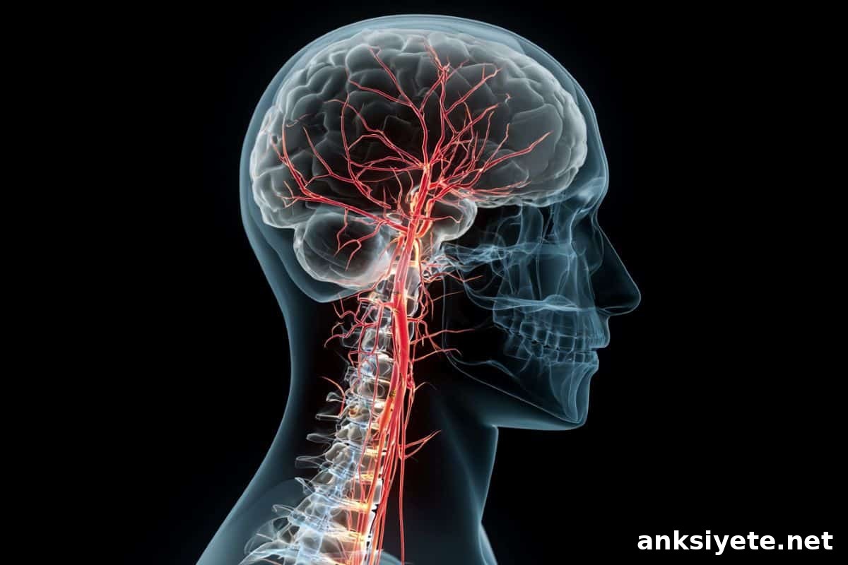
“Our study shows, at the cellular level, how the central nervous system adapts its control of movement after incomplete SCI,” says Ruoli Wang, associate professor in biomechanics at the Promobilia MoveAbility lab, KTH Royal Institute of Technology. The team emphasized that all measurements were non-invasive.
The peer-reviewed paper was published in the Journal of NeuroEngineering and Rehabilitation.
Lead author Zhihao Duan reports that, after injury, the nervous system struggles to distribute coordinated signals across motor units during low-force tasks. As force demands increase, the system shifts into a less nuanced mode, producing stronger but less flexible drive that appears to prioritize gross force over precision.
A single muscle contracts by recruiting and modulating hundreds of motor units—each comprising a motor neuron and the muscle fibers it innervates. When motor units in different muscles receive a common neural input they can act together as a coordinated group, similar to sections in an orchestra led by a conductor. That shared input is essential for smooth, adaptable movement.
To assess coordination, the researchers examined 25 participants in total—fifteen people with incomplete SCI and ten non-disabled controls. They recorded HD-EMG signals from the soleus and gastrocnemius medialis while participants produced isometric plantarflexion at two target force levels: 20% and 50% of maximal voluntary contraction. The HD-EMG signals were decomposed into individual motor unit spike trains, and analyses included inter-muscle coherence to quantify shared neural drive and factor analysis to identify motor unit clusters.
At the lower 20% effort level, the SCI group showed a reduced proportion of motor units in shared clusters across the two calf muscles, indicating fewer units driven by the same coordinated signal. Clinically, this reduced shared drive corresponds to shaky, unstable low-force control. At the higher 50% effort level, SCI participants exhibited increased low-frequency inter-muscle coherence, consistent with a stronger but less differentiated neural drive that produces rigid, over-synchronized muscle activation.
“One striking outcome is that after SCI the nervous system appears less able to change strategy as force increases,” Wang explains. “A healthy system can scale and alter the shared neural drive to match task demands; the SCI-affected system becomes more rigid and less adaptive.”
The authors note limitations, including the modest sample size and the technical challenge of identifying many motor units from surface recordings. Despite these constraints, the findings provide a new view of how SCI reshapes motor unit synergies and suggest measurable biomarkers that could inform targeted neurorehabilitation approaches designed to restore coordinated neural input.
Funding: The study was a collaboration with Aleris Rehab Station and received funding from the Swedish Research Council and the Promobilia Foundation.
Key Questions Answered:
A: Walking relies on momentum and larger, coordinated muscle actions. Standing still or balancing demands low-level, precise coordination of many small motor units. The study found that at low effort (about 20% of maximum), the shared neural drive across motor units is weakened after SCI, so fine-tuned stability is compromised.
A: The term describes a change in how the nervous system encodes command strength. Instead of finely graded, selective inputs, the injured system appears to send broader, higher-amplitude signals when more force is required. That “louder” drive recruits units in a more uniform, less differentiated way, producing rigid rather than precise movements.
A: Rehabilitation often emphasizes strength training. These results suggest therapies should also target the neural coordination—the timing and shared drive among motor units. Objective electrical biomarkers from HD-EMG could help clinicians design interventions, exercises, or stimulation programs aimed at re‑establishing a flexible, coordinated neural drive.
Editorial Notes:
- This article was edited by a Neuroscience News editor.
- The journal paper was reviewed in full by editorial staff.
- Additional explanatory context was provided by the editorial team.
About this SCI and neurology research news
Author: David Callahan
Source: KTH Royal Institute of Technology
Contact: David Callahan – KTH
Image: Image credited to Neuroscience News
Original Research: Open access. “Adaptation of motor unit synergies in the synergetic ankle plantarflexors in ambulatory persons with incomplete spinal cord injury” by Zhihao Duan, Asta Kizyte, Emelie Butler Forslund, Elena M. Gutierrez-Farewik, Pawel Herman & Ruoli Wang. DOI: 10.1186/s12984-026-01874-2
Abstract
Adaptation of motor unit synergies in the synergetic ankle plantarflexors in ambulatory persons with incomplete spinal cord injury
Background
Spinal cord injury commonly impairs motor coordination. Prior work has shown that muscle synergies help coordinate movement and that these synergies change after SCI. However, how adaptations occur at the motor unit level—where neural commands meet muscle fibers—has not been fully explored. This study investigated motor unit synergies and clustering in the soleus and gastrocnemius medialis muscles and examined how these patterns differ in people with incomplete SCI.
Methods
High-density electromyography (HD-EMG) recorded motor unit activity in the soleus and gastrocnemius medialis muscles of fifteen participants with incomplete SCI and ten non-disabled participants. Recordings were taken during isometric contractions at 20% and 50% of maximal voluntary force. Signals were decomposed into individual motor unit spike trains. Inter-muscle coherence evaluated shared neural drive, and factor analysis identified synergistic motor unit clusters within each muscle.
Results
Both groups exhibited overall shared neural drive between the soleus and gastrocnemius medialis, supporting coordinated plantarflexor function. Compared with controls, participants with SCI showed altered coherence in the delta frequency band, with significantly higher low-frequency coherence at 50% maximal voluntary contraction (p = 0.047). Factor analysis also revealed a reduced proportion of motor units in the shared cluster within the gastrocnemius medialis at 20% maximal voluntary contraction (p < 0.01) in the SCI group.
Conclusions
The findings indicate that incomplete SCI can disrupt motor unit synergies and clustering, which may undermine coordinated motor control. These insights clarify neural adaptations after SCI and provide a foundation for developing rehabilitation strategies that aim to restore more adaptive, coordinated neural drive.