
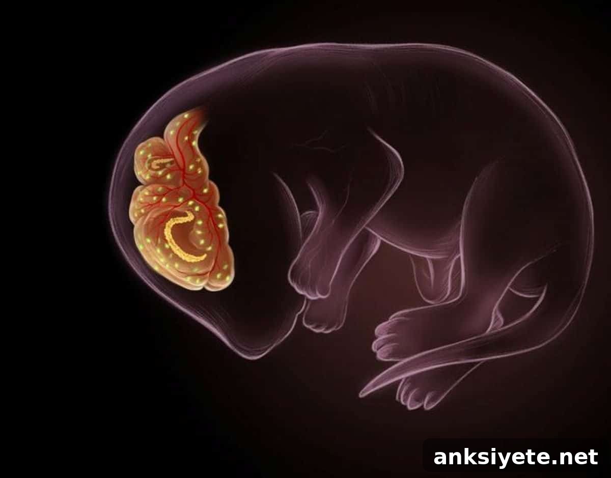
Summary: A new study overturns the long-standing view of senescent cells as solely damaging “zombie” cells. Researchers found that, during mouse embryonic development, certain non-dividing senescent cells appear at precise times to guide the formation of the brain’s two main protective interfaces: the blood-brain barrier (BBB) and the blood–cerebrospinal fluid (CSF) barrier. While most developmental senescence is transient, the team discovered that choroid plexus epithelial cells retain senescent characteristics into adulthood, revealing that senescence can be a purposeful, context-dependent process essential for brain structure and function.
Using single-cell RNA sequencing, genetic lineage tracing, and high-resolution imaging in mouse embryos, investigators identified three distinct cell types that enter senescence temporarily to coordinate vascular patterning, fluid balance, and barrier integrity. When these senescent cells were selectively removed during gestation, embryos developed severe defects in barrier formation and intracranial fluid homeostasis, demonstrating that these cells are functionally required for healthy brain development.
Key Facts
- Paradigm shift: Senescence is not exclusively a marker of aging or disease; it is also essential for key stages of embryonic brain development.
- Barrier architects: Specialized senescent cells appear during development to build the blood-brain and blood-CSF barriers; their absence produces structural and fluid-balance defects.
- Three specialized cell types: Vascular endothelial cells, brain-resident macrophages, and choroid plexus epithelial cells transiently or persistently adopt senescent states to support barrier formation.
- Transient versus persistent: Endothelial and macrophage senescence is temporary and tied to vascular patterning, whereas a subset of choroid plexus epithelial cells maintains senescent features into adulthood.
- Coordinated signaling hubs: Senescent cells act as communicative centers, coordinating different cell types to assemble vascular and epithelial defenses rather than simply being isolated, dying units.
- Implications for disease: The finding that senescence assumes diverse molecular states depending on cell type and developmental timing suggests new avenues for studying how these pathways might malfunction in adult brain disorders.
Source: UCSD
The brain’s protective barriers
The blood-brain and blood-CSF barriers are specialized cellular interfaces that allow nutrients to reach neural tissue while preventing toxins and pathogens in the circulation from entering the central nervous system. Although their functions have been well characterized, the cellular processes that build these barriers during development have been less clear—until now.
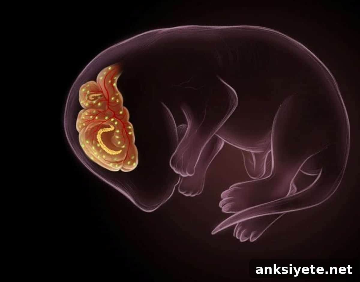
Led by researchers at the University of California San Diego and published in Cell, this study identifies developmentally programmed p21+ senescent cells that adopt distinct, context-dependent roles at two brain interfaces. In the choroid plexus, epithelial cells take on a lifelong, non-inflammatory senescent state associated with CSF production and blood–CSF barrier integrity. By contrast, vascular endothelial cells and brain-resident macrophages display transient, pro-inflammatory senescence profiles during vascular growth, coordinating angiogenic patterning and extracellular matrix assembly.
The research team combined single-cell transcriptomics, lineage tracing, and imaging to map when and where senescent states arise. They observed that endothelial and macrophage senescence appears during periods of vessel remodeling and then resolves after blood vessels are patterned. Choroid plexus epithelial cells were unusual in retaining senescence-associated features long after development, persisting into adult life without evident inflammatory consequences.
To test the functional importance of these states, investigators used genetic approaches to ablate p21+ cells during mid-gestation. Embryos lacking these developmental senescent cells displayed disrupted brain vascular patterning, hemorrhage, impaired CSF production, ventricular collapse, and overall failure to maintain normal fluid balance—strong evidence that senescent cells actively shape developing brain barriers.
“We were surprised to find that senescence presents very differently depending on cell type and developmental timing,” said the study’s lead scientists. “These cells do not act in isolation; they form coordinated signaling networks that guide barrier assembly and, in the case of the choroid plexus, help sustain barrier function into adulthood.”
Key Questions Answered
A: Timing and persistence matter. Transient developmental senescence helps coordinate tissue assembly and is typically resolved or cleared. In contrast, senescent cells that accumulate in aged tissues often persist and adopt a pro-inflammatory secretory profile (SASP) that harms neighboring cells. Senolytic strategies target these pathological, lingering adult cells rather than the beneficial, tightly regulated embryonic senescent states.
A: The team used genetic models to selectively eliminate p21+ senescent cells during mouse gestation. Embryos without these cells developed malformed blood vessels, disrupted barrier architecture, and severe disturbances in CSF production and intracranial pressure—showing these cells are necessary for normal barrier formation.
A: Developmental senescence has generally been categorized as temporary. Discovering a population of choroid plexus cells that maintain a senescent signature from embryogenesis into adulthood challenges that assumption and suggests senescence can be a stable, functional state that supports ongoing organ function.
Editorial Notes
- This article was edited by a Neuroscience News editor.
- The full journal paper was reviewed.
- Additional explanatory context was added by staff for clarity.
About this neuroscience research news
Author: Mario Aguilera
Source: UCSD
Contact: Mario Aguilera – UCSD
Image: Credit to Ella Maru Studio; conceptualized by Ashley Watson and Hiruy Meharena
Original Research: Open access. “Persistent and transient senescent cells contribute to brain barrier development” by L. Ashley Watson, Zoe Adelsheim, Mackenzie J. Carter, Grace T. Carter, Karen L. Jimenez-Reyes, Huijie Du, Ziqing Zhu, David B. Berry, Mia C. Paredez, Rania H. Palaniappan, John M. Augustine, and Hiruy S. Meharena. Published in Cell.
Abstract
Persistent and transient senescent cells contribute to brain barrier development
The establishment of the blood-brain barrier and the blood–CSF barrier requires precise coordination among diverse cell types to protect and nourish the developing brain. This study identifies p21+ senescent cells programmed during development that display divergent senescence-associated features at the two interfaces in mice. In the choroid plexus, epithelial cells adopt a long-lived, non-inflammatory senescent state linked to CSF production and blood–CSF barrier integrity. In contrast, vascular endothelial cells and brain-resident macrophages transiently present pro-inflammatory senescence signatures during vascularization, engaging in reciprocal signaling that supports angiogenic patterning and extracellular matrix assembly. Genetic ablation of p21+ cells during mid-gestation disrupts vascular patterning and choroid plexus integrity, causing hemorrhage, impaired CSF production, and ventricular collapse. These results indicate that embryonic senescent cells can adopt transient or long-lived states that support brain-barrier formation and homeostasis, reframing persistent senescence as not solely pathological but sometimes an adaptive, functional program.