
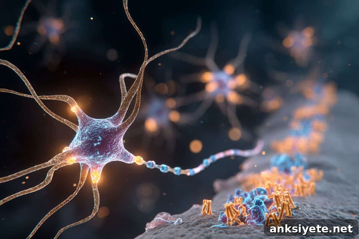
Summary: New research shows that neurons release an extracellular kinase called VLK that strengthens synapses by phosphorylating receptors outside the cell. VLK phosphorylates EphB receptors on the postsynaptic membrane, which promotes recruitment and clustering of N-methyl-D-aspartate (NMDA) receptors and thereby amplifies synaptic signaling. This mechanism fills a key gap in understanding how the nervous system adapts during learning and how pain hypersensitivity develops after injury.
In mice, deleting VLK reduced pain hypersensitivity after injury, while applying VLK increased NMDA-dependent activity in both mouse and human neurons. These findings change long-held assumptions about where kinase activity can act and point to a potential way to influence plasticity without directly altering the central signaling functions of NMDA receptors.
Key Facts:
- Extracellular kinase function: VLK functions within the synaptic cleft to phosphorylate postsynaptic receptors and enhance NMDA receptor clustering.
- New pain mechanism: Removing VLK prevents injury-induced pain hypersensitivity, identifying a novel molecular target for possible analgesic strategies.
- Wider neuroscience impact: VLK-driven modulation of synapses may affect learning, memory, and plasticity across multiple brain regions.
Source: UT Dallas
Researchers at The University of Texas at Dallas’ Center for Advanced Pain Studies (CAPS) and collaborators have identified a critical mechanism by which synaptic connections become stronger.
The discovery, reported Nov. 20 in the journal Science, has direct implications for understanding the biochemical processes underlying learning, memory and pain, said Dr. Ted Price BS’97, Ashbel Smith Professor of neuroscience in the School of Behavioral and Brain Sciences, CAPS director and co-corresponding author of the study.
“This study addresses the core mechanisms of synaptic plasticity — how neural connections change,” Price said. “Its implications extend across neuroscience.”
The team’s work centers on phosphorylation, the biochemical addition of a phosphate group to a protein by an enzyme called a kinase. Phosphorylation is a well-established regulator of intracellular processes such as metabolism and signaling. By contrast, phosphorylation that occurs outside the cell — extracellular phosphorylation — has been poorly understood, especially at synapses.
Synapses are the spaces where presynaptic neurons release neurotransmitters, peptides and proteins that bind and regulate receptors on postsynaptic cells. These exchanges underlie synaptic plasticity, the process that strengthens or weakens connections and supports learning, memory and pain signaling.
The researchers investigated whether kinases secreted by neurons, called ectokinases, control synaptic signaling by phosphorylating receptors in the synaptic cleft.
“Extracellular phosphorylation is carried out by ectokinases — kinases secreted outside cells. It’s been recognized for a long time, but its role in the nervous system has been largely unexplored,” Price said. “Our study shows that kinases released into the synaptic cleft directly influence synaptic plasticity, updating textbook ideas about how synapses function.”
Previous studies linked extracellular phosphorylation to pain, but the responsible kinase was unknown. The team focused on vertebrate lonesome kinase (VLK, gene name Pkdcc), previously implicated in platelet function and bone development. Their experiments indicate VLK is released by presynaptic neurons after injury and phosphorylates the extracellular domain of ephrin type-B receptor 2 (EphB2) on postsynaptic membranes.
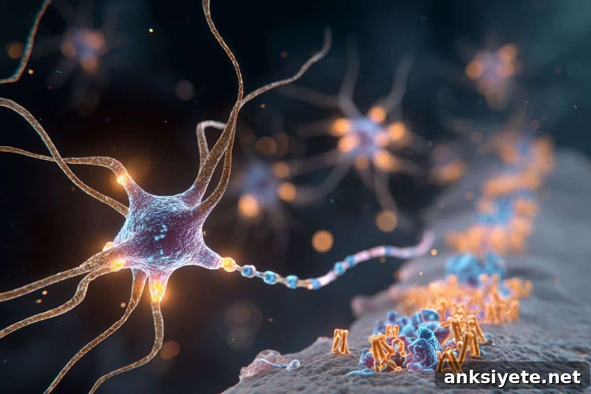
Phosphorylation of EphB2 attracts NMDA receptor proteins, which then cluster with EphB2 at the membrane. NMDA receptors are central to learning and memory because they regulate neuronal electrical responses that strengthen synaptic connections.
“Increasing NMDA receptor concentration at the synapse permits stronger neuronal activation and larger postsynaptic potentials — a core mechanism of synaptic plasticity,” said Hajira Elahi BS’17, MS’19, PhD’23, a co-first author who conducted much of the work as part of her dissertation.
Mice engineered to lack VLK in sensory neurons involved in pain did not develop acute mechanical hypersensitivity after surgical injury. In contrast, administering recombinant VLK to normal mice produced strong pain hypersensitivity that depended on NMDA receptor activation.
“We also found that human sensory neurons express and secrete VLK, and that VLK promotes the EphB2–NMDA receptor interaction in human tissue,” Elahi added. “This underlines the translational relevance of our findings.”
Price noted the project began a decade ago through collaboration with the Dalva lab. “After years of work, it became clear that VLK phosphorylates EphB1 and EphB2 and that VLK activity is sufficient to cause NMDA receptor clustering,” he said.
Dr. Matthew Dalva, co-corresponding author and director of the Tulane Brain Institute, emphasized the broader significance: “Neurons releasing a protein kinase to modify synaptic function suggests many new and unexpected targets for research and therapy. This project exemplifies collaborative science; our findings depended on expertise from multiple teams.”
Directly targeting NMDA receptors for pain relief has proven difficult due to widespread roles of these receptors across the nervous system and associated side effects. By contrast, targeting VLK offers a new route to modulate NMDA receptor function indirectly and potentially more selectively. In cortical neurons, VLK release appears to be activity dependent, suggesting spatially and temporally specific control of receptor function.
A prospective therapy could involve locally blocking VLK in the spinal cord to reduce pathological pain, though more work is required to determine how broadly this synaptic VLK mechanism operates throughout the nervous system.
“Discovering that kinases act within the synaptic cleft — not only inside cells — is a major advance in understanding how receptors that drive synaptic plasticity are regulated,” Price said. “We have likely only begun to uncover the roles of extracellular kinase signaling at synapses.”
UT Dallas-affiliated authors include neuroscience research scientists Moeno Kume BS’17 PhD’23, Ishwarya Sankaranarayanan PhD’22 and Stephanie Shiers PhD’19; doctoral students Lucy He and Khadijah Mazhar; and Juliet Mwirigi BS’17, PhD’23, Jessica Loucks BS’22 and Rohita Arjarapu BS’24. Additional contributors are from Tulane, UT San Antonio Health Science Center, UT MD Anderson Cancer Center, University of Houston Tilman J. Fertitta Family College of Medicine, Princeton University, University of Wisconsin-Madison, NYU Grossman School of Medicine and Thomas Jefferson University.
Funding: This research was supported by grants from the National Institute of Neurological Disorders and Stroke (R01NS111976, R01NS115441, U19NS130608), the National Institute on Drug Abuse (R01DA022727) and the National Center for Research Resources (S10RR027990), all parts of the National Institutes of Health.
Key Questions Answered:
A: VLK phosphorylates extracellular sites on EphB receptors, which triggers NMDA receptor clustering and strengthens synaptic connections.
A: The VLK-driven mechanism enhances synaptic signaling after injury, clarifying processes that underlie memory formation and injury-induced pain hypersensitivity.
A: Potentially. Blocking VLK could reduce pathological pain while avoiding the widespread side effects associated with direct NMDA receptor inhibition.
Editorial Notes:
- This article was edited by a Neuroscience News editor.
- The journal paper was reviewed in full.
- Additional context was added by the editorial staff.
About this synaptic plasticity research news
Author: Stephen Fontenot
Source:UT Dallas
Contact: Stephen Fontenot – UT Dallas
Image: The image is credited to Neuroscience News
Original Research: Closed access.
“The synaptic ectokinase VLK triggers the EphB2–NMDAR interaction to drive injury-induced pain” by Ted Price et al. Science
Abstract
The synaptic ectokinase VLK triggers the EphB2–NMDAR interaction to drive injury-induced pain
INTRODUCTION
Protein phosphorylation is a fundamental mechanism of intracellular regulation, but phosphorylation outside the cell has been less well characterized. Although extracellular phosphorylation of secreted proteins like casein has been known since the 19th century, its physiological roles at synapses have remained unclear. Recent work has identified secreted kinases and shown that many synaptic proteins carry phosphorylated extracellular domains, but whether extracellular phosphorylation alters synaptic signaling or behavior in vivo was previously unresolved.
RATIONALE
The study set out to determine whether extracellular phosphorylation regulates receptor interactions at synapses. Specifically, the researchers tested whether a secreted kinase could control interaction between the receptor tyrosine kinase EphB2 and the NMDA receptor (NMDAR), a central regulator of glutamate signaling and pain. Because phosphorylation of EphB2 at the conserved extracellular residue Y504 is required for NMDAR binding, the team asked whether secreted tyrosine kinases could mediate this modification and thereby influence pain-related behaviors in vivo. Their aim was to map a synaptic signaling pathway linking presynaptic activity to extracellular receptor phosphorylation and downstream NMDAR function.
RESULTS
The investigators identified vertebrate lonesome kinase (VLK/Pkdcc) as the single member of a six-gene ectokinase family that is both secreted and sufficient to induce EphB2–NMDAR interaction. VLK phosphorylated EphB2 at Y504 in an ATP-dependent manner; the effect was blocked by extracellular phosphatase and absent with kinase-dead VLK mutants. Recombinant VLK induced EphB2–NMDAR complex formation in cultured neurons and spinal tissue, while genetic knockout of Pkdcc abolished the interaction. VLK localized to synaptic vesicles and was released in a SNARE-dependent manner following ephrin-B stimulation or increased neuronal activity, consistent with regulated synaptic secretion.
In vivo, conditional deletion of Pkdcc from presynaptic sensory neurons prevented EphB2 phosphorylation and blocked EphB2–NMDAR interaction in dorsal horn projection neurons after injury, without changing baseline protein expression. Mice lacking VLK in sensory neurons did not develop mechanical hypersensitivity after surgical injury but maintained normal motor coordination and normal responses to heat and chemical stimuli. Intrathecal injection of recombinant VLK induced EphB2–NMDAR interaction and pain-like behaviors; this effect required NMDAR activity. Deleting EphB2 postsynaptically in spinothalamic neurons prevented injury-induced pain behaviors, confirming the necessity of this pathway for pain sensitization.
CONCLUSION
These results reveal an extracellular phosphorylation pathway that links presynaptic activity to postsynaptic receptor organization and behavior. By releasing a kinase that acts on the outside of the postsynaptic membrane, neurons can modulate NMDAR function in a spatially and behaviorally specific way, independent of canonical intracellular signaling. Given the essential role of NMDARs in synaptic plasticity and the developmental consequences of global neuronal VLK loss, this pathway likely extends beyond pain and may open new therapeutic possibilities for tuning receptor function.