
Summary: Researchers have uncovered how the brain protein cypin preserves strong connections between neurons, a process essential for learning and memory. Cypin controls how proteins are tagged and positioned at synapses, enabling reliable neural communication and supporting cognitive function.
The study also shows that cypin slows the breakdown of certain proteins and increases levels of synaptic proteins, strengthening signaling between brain cells. These discoveries identify cypin as a promising target for therapies to repair brain injury and counteract neurodegenerative disease.
Key Facts:
- Cypin helps add and position molecular tags on synaptic proteins, supporting efficient neuron-to-neuron communication.
- It interacts with the proteasome system to reduce protein degradation, raising levels of key synaptic components.
- Boosting cypin activity could help preserve memory and learning, and may offer new treatment strategies for brain injury and diseases like Alzheimer’s and Parkinson’s.
Source: Rutgers University
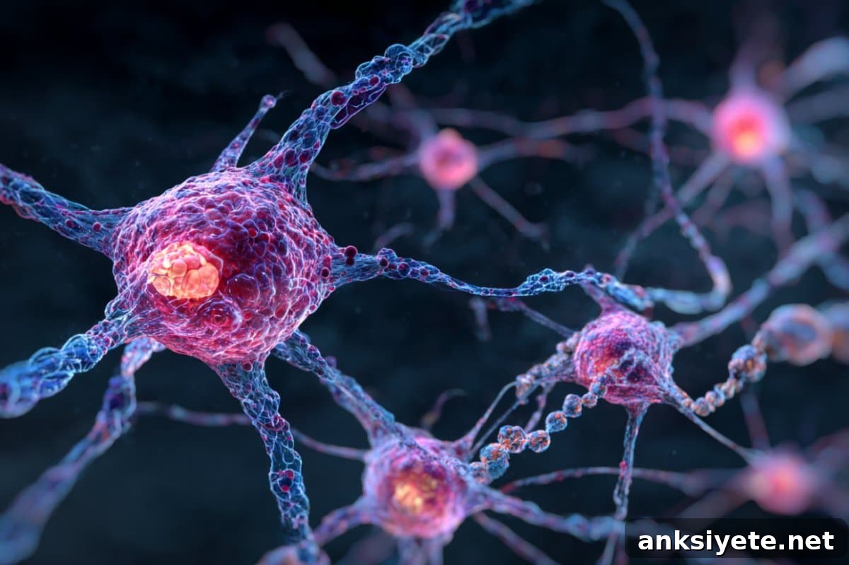
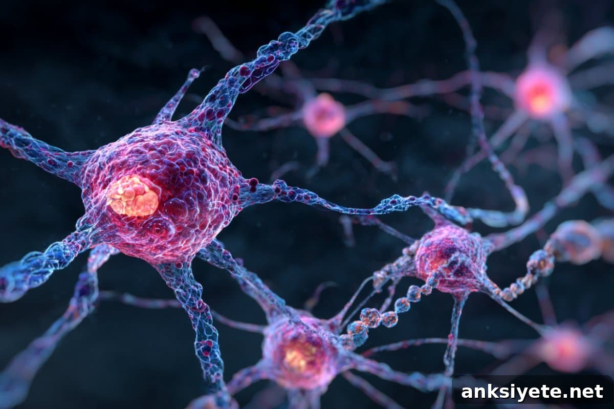
Researchers have identified a critical role for cypin in maintaining the molecular architecture of synapses, the specialized junctions where neurons exchange information.
Published in Science Advances, the work—led by a Rutgers University–New Brunswick laboratory—reveals that cypin shapes synaptic content by promoting specific molecular tags on proteins at synapses. Those tags help ensure the right proteins are present in the right place so synapses can transmit signals reliably.
Bonnie Firestein, a Distinguished Professor in the Department of Cell Biology and Neuroscience, and colleagues reported that cypin helps add K63-linked ubiquitin tags to postsynaptic proteins. These modifications influence where proteins end up at synapses and how they function, a key factor for sustained neuronal communication and plasticity.
Beyond tagging, the team found that cypin interacts with the proteasome—the cellular machinery that normally breaks down proteins—and by doing so it slows degradation of certain synaptic proteins. Slower turnover increases the abundance of these proteins at synapses, which supports stronger signaling between neurons.
Firestein’s experiments also show that raising cypin levels increases synaptic protein content and enhances related signaling processes tied to learning and memory. The protein further appears to amplify the activity of UBE4A, an enzyme that contributes to the ubiquitin-tagging process, suggesting a coordinated mechanism by which cypin influences synaptic composition.
“Although this work is foundational biology, it points toward realistic therapeutic pathways,” Firestein said. Her lab is pursuing translational studies that could eventually convert these molecular insights into clinical approaches for improving cognitive function after injury or during disease.
Healthy synaptic function is often damaged in conditions such as Alzheimer’s and Parkinson’s disease and following traumatic brain injury. Because cypin supports both the placement of synaptic proteins and synaptic plasticity—the capacity of synapses to adapt and remodel over time—modulating cypin could help restore or preserve synaptic health in those conditions.
The study combined experiments in cultured neurons with in vivo work in adult mice, giving the findings broader relevance across developmental stages and biological contexts. By linking cypin to K63-linked polyubiquitination and to proteasome interaction, the research adds an important layer of understanding to how synaptic protein composition is regulated.
Funding: This research was supported in part by the National Institutes of Health (NINDS), the Coalition for Brain Injury Research, a charitable foundation honoring the memory of Dennis John Benigno, and private donors Jamuna Rajasingham and Dyan Rajasingham.
Collaborators from Rutgers include Kiran Madura of the Department of Pharmacology at Robert Wood Johnson Medical School and former doctoral students Srinivasa Gandu, Mihir Patel and Ana Rodriguez in the Department of Cell Biology and Neuroscience. Jared Lamp and Irving Vega of Michigan State University also contributed to the project.
About this genetics and memory research news
Author: Kitta MacPherson
Source: Rutgers University
Contact: Kitta MacPherson – Rutgers University
Image: The image is credited to Neuroscience News
Original Research: Open access.
“Cypin regulates K63-linked polyubiquitination to shape synaptic content” by Bonnie Firestein et al. Science Advances
Abstract
Cypin regulates K63-linked polyubiquitination to shape synaptic content
A central question in neuroscience is how posttranslational modifications of proteins determine their localization and function at synapses. Polyubiquitination is known for directing proteins to the proteasome for turnover, but distinct ubiquitin linkages can have other signaling roles.
K63-linked polyubiquitin (K63-polyUb) has been studied in non-neuronal contexts such as signal transduction in cancer, yet its neuronal functions remain less defined. Here, we identify a role for the cytosolic PSD-95 interactor cypin in promoting K63-polyUb on postsynaptic proteins and shaping synaptic content.
Using in vitro neuronal development models and in vivo analysis in adult mice, we show that cypin promotes K63-linked polyubiquitination on synaptic proteins and exerts effects on both postsynaptic and presynaptic function. These cypin-driven modifications influence synaptic composition and signaling, providing new insight into mechanisms that regulate neuronal communication.