
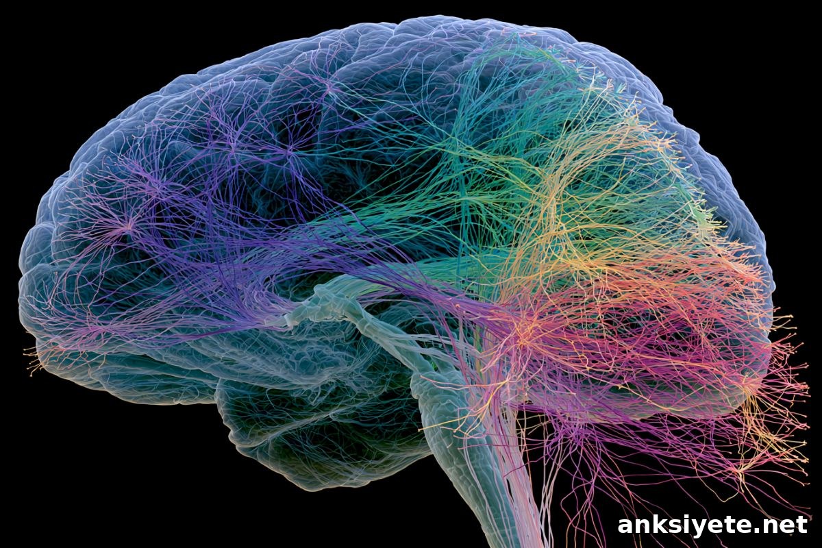
Summary: A collaborative study combines detailed rodent circuit mapping with ultra-high-field 7T human fMRI to reveal how upstream brain regions regulate the periaqueductal gray (PAG), a core midbrain center for defensive responses. By identifying the human networks that inhibit or modulate the PAG, the team aims to develop real-time fMRI neurofeedback therapies that help people with PTSD and anxiety disorders consciously reduce persistent hyper-arousal.
Key Facts
- The Periaqueductal Gray (PAG) Epicenter: The PAG is a compact midbrain structure surrounding the cerebral aqueduct. It serves as a central command node for survival behaviors—fight, flight, or freeze. In many individuals with PTSD and anxiety disorders, PAG-driven defensive activity can remain abnormally elevated because the normal regulatory mechanisms fail to suppress it.
- Indirect Targeting Strategy: Because the PAG is very small and deeply situated in the brainstem, it is difficult to target directly with noninvasive therapies. The research therefore focuses on the larger cortical and subcortical regions that exert inhibitory control over the PAG. These upstream regulators are more accessible targets for neurofeedback or other interventions.
- Rodent-to-Human Translation Loop:
- Amsterdam (NIN): Using precise tools such as optogenetics and calcium imaging in mice, researchers map the specific cell types and circuits that enable fear extinction and stress control.
- Maastricht (UMC): Using ultra-high-field 7 Tesla fMRI, human imaging teams test whether the same circuits identified in rodents are structurally and functionally present in people, validating translational relevance.
- Real-Time Neurofeedback: The long-term goal is to create individualized fMRI neurofeedback protocols. Patients in a 7T scanner would receive continuous visual feedback of activity in their upstream threat-regulation networks, learning over repeated sessions to downregulate overactive responses and strengthen inhibitory control over the PAG.
- Educational Mission: Beyond therapy development, the collaboration is integrating these circuit-level findings into training programs for psychiatrists, psychologists, and clinical researchers, so future clinicians understand the neural wiring that underlies fear and stress regulation.
Source: KNAW
Why does the brain’s stress system remain stuck in overdrive for some people?
Scientists at the Netherlands Institute for Neuroscience (NIN) in Amsterdam and Maastricht UMC are working together to answer this question. By combining mechanistic animal research with high-resolution human imaging, they seek to clarify how regulatory brain networks fail to suppress the PAG in stress-related disorders and to translate those insights into targeted interventions for PTSD and anxiety.
The brain’s alarm system
The body’s automatic response to perceived danger is familiar: increased heart rate, muscle tension, and rapid mobilization for action. For some people with stress-related conditions, that alarm system remains active even when threats have passed. Central to that process is the periaqueductal gray (PAG), a compact midbrain structure that coordinates immediate defensive responses. Neurobiologist Alexander Heimel explains that while the PAG executes survival behaviors, higher-level brain areas normally provide inhibitory control. When those braking signals fail, the PAG can drive chronic hyper-vigilance.
From the laboratory to the clinic
The project uses two complementary approaches. In Amsterdam, detailed experiments in rodents identify the neuronal cell types and synaptic pathways that enable fear learning and extinction. These findings generate precise hypotheses about which upstream regions modulate the PAG. In Maastricht, researchers test these hypotheses in humans using 7 Tesla functional MRI, which offers the spatial resolution needed to observe activity patterns near the brainstem. If the implicated networks are present and measurable in people, the team will develop neurofeedback protocols that train participants to alter activity in those networks.
During neurofeedback training, participants view a simple visual representation—such as a moving bar or thermometer—that reflects activity in their own stress-regulation network. Guided by cognitive strategies and repeated practice, people learn to reduce that signal. Over sessions, this learning aims to strengthen the brain’s inhibitory control and decrease pathological threat responses.
A unique collaboration
What sets this initiative apart is the close integration between basic neuroscience and clinical psychiatry. Heimel notes that relatively few projects link animal circuit discovery so tightly with human imaging and potential clinical applications. Psychiatrist David Linden emphasizes that rigorous knowledge about the networks to be influenced is a prerequisite for developing effective treatments. By aligning mechanistic discovery with translational testing from the outset, the collaboration hopes to accelerate the path from bench to bedside.
The team also prioritizes education: they plan to include the circuit-level insights in curricula for early-career mental health professionals so clinicians will better understand the neural foundations of fear and stress, which in turn can inform therapeutic decisions and innovation.
Key Questions Answered:
A: The PAG is an evolutionarily ancient midbrain structure that coordinates immediate survival responses such as freezing, fleeing, or fighting. In healthy brains, cortical and subcortical regions regulate PAG activity. In PTSD, those regulatory systems can fail, allowing the PAG to remain overactive and contributing to persistent hyper-vigilance and distress.
A: Standard clinical scanners usually operate at 1.5T or 3T and provide good anatomical images but lack the spatial precision needed to resolve tiny structures near the brainstem. Ultra-high-field 7T fMRI provides much finer spatial resolution, enabling researchers to observe activity and connectivity patterns in small, deep regions like the PAG and its upstream regulators in awake human participants.
A: Neurofeedback acts as a real-time mirror of brain activity. While inside an fMRI scanner, individuals see a visual indicator that reflects activity in their stress-regulation network. With guidance and practice, they discover mental strategies that lower that signal. Repeated training can strengthen inhibitory pathways and help the brain adopt a calmer response pattern to perceived threats.
Editorial Notes:
- This article was edited by a Neuroscience News editor.
- Journal paper reviewed in full.
- Additional context added by our staff.
About this PTSD and neuroscience research news
Author: Eline Feenstra
Source: KNAW
Contact: Eline Feenstra – KNAW
Image: The image is credited to Neuroscience News