
Summary: A large brain imaging study led by researchers at the University of Kansas finds a strong association between lifetime exposure to reproductive hormones and structural brain health in older women.
The study analyzed 459 women aged 65 to 80 and found that greater cumulative exposure to estrogen — from early-life hormonal contraceptives, menopausal hormone therapy (MHT) in midlife, or a naturally later menopause — was linked to larger brain volumes and thicker cortex in regions important for memory, information processing and higher cognitive functions.
Key Findings
- Lifespan perspective: Use of estrogen-containing oral contraceptives in young adulthood leaves a detectable, protective structural signature in the brain decades later.
- Neuroprotective roles of estrogen: Estrogen supports white matter integrity, helps preserve neurons, strengthens synaptic connections and contributes to healthy cerebral vascular function.
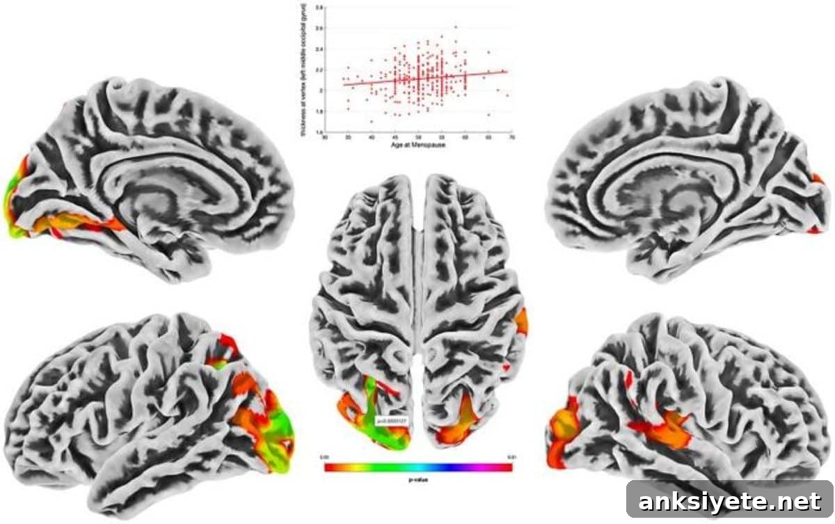
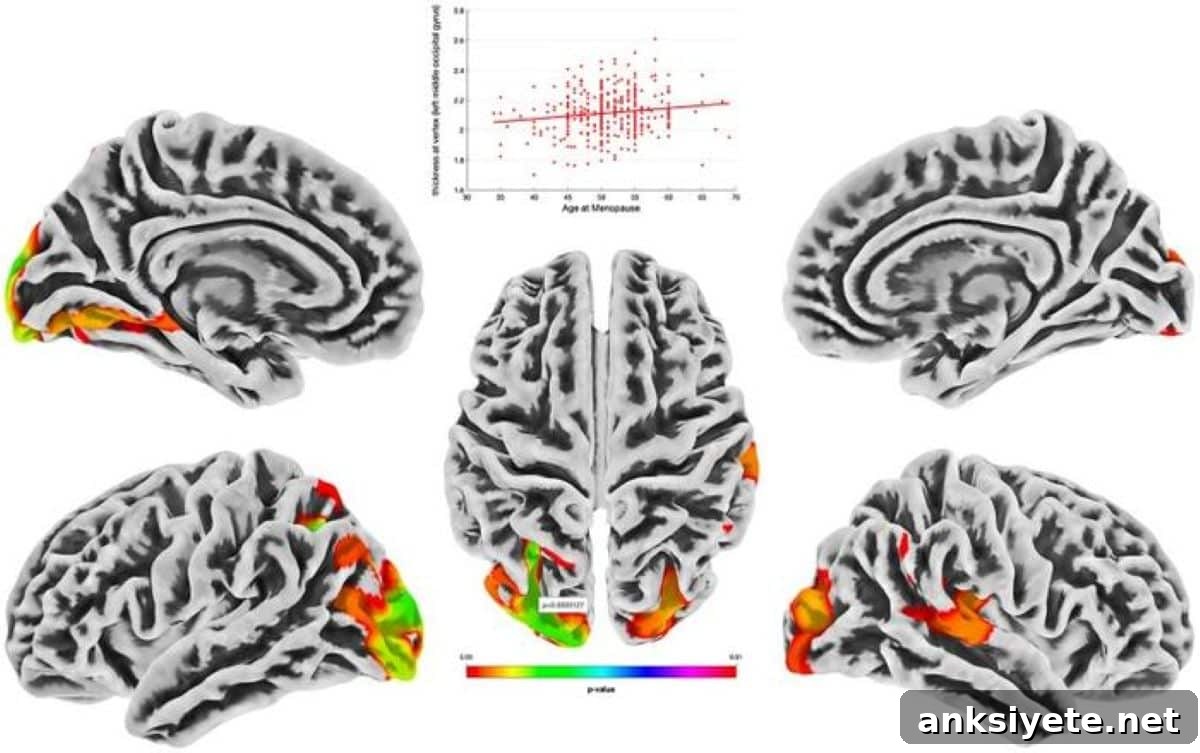
- Later menopause: Women with later natural menopause — indicating longer endogenous ovarian hormone exposure — showed thicker cortex in brain regions that are often vulnerable in Alzheimer’s disease.
- Benefits after surgical menopause: Women who had early ovarian removal (oophorectomy) showed marked structural brain benefits when they received estrogen replacement.
- Reassessing past concerns: These results contribute to the re-evaluation of conclusions drawn from the 2002 Women’s Health Initiative (WHI) study, underscoring that hormone therapy effects depend on timing, population and study design.
Source: University of Kansas
Overview of the study
Researchers at the University of Kansas examined structural MRI scans and retrospective reports of hormonal medication use in 459 women aged 65–80 enrolled in the IGNITE study. The analysis considered three sources of lifetime ovarian hormone exposure: hormonal birth control (BC) during early adulthood, menopausal hormone therapy (MHT) during midlife, and endogenous exposure estimated by age at natural menopause.
“Our results indicate a protective effect: lifetime exposure to estrogen-based therapies is associated with structural features of brain health in older women,” said co-lead author Amber Watts, professor of psychology at KU. The team looked at both early- and midlife hormone medication use and found each contributed to brain measures decades later.
The investigators propose that the cumulative duration of estrogen exposure — beginning with oral contraceptive use in young adulthood and extending through menopausal therapy when applicable — may underlie these positive associations.
The study grouped exposures into three categories:
- Use of hormonal birth control (BC) in early adulthood
- Use of menopausal hormone therapy (MHT) in midlife
- Natural hormone exposure measured by age at menopause
Watts emphasized that most prior research has treated menopause as the primary hormonal event of interest. This study expands that view by considering how earlier hormonal exposures — occurring while the brain is still developing — may influence structural aging decades later.
Given that a large proportion of women of reproductive age use hormonal contraception, these findings have potential long-term relevance for individual and clinical decision-making about reproductive and menopause-related therapies.
“Estrogen is more than a reproductive hormone,” Watts said. “It supports multiple body systems, including the brain, heart, bones and immune system. In the brain it can protect white matter, maintain neuronal health, support synaptic connections and preserve vascular function.”
The team also observed that women with later natural menopause tended to have greater cortical thickness in posterior regions commonly affected by Alzheimer’s disease, reinforcing the idea that longer endogenous hormone exposure may relate to preserved brain structure.
Regarding the controversy around hormone therapy: the authors note that the 2002 Women’s Health Initiative raised concerns that reduced the use of estrogen therapies for many years. Subsequent work has revealed limitations in those early findings. The current study aligns with accumulating evidence that, when appropriately prescribed, estrogen therapies can have beneficial effects on aging systems including the brain.
The new investigation complements Watts’ prior work linking birth control and menopausal hormone use to cognitive outcomes and further highlights benefits for women who received hormone therapy after early surgical menopause.
Watts and colleagues continue to recruit participants for additional menopause-related studies and encourage interested women to inquire about study eligibility.
Lead investigators include Robyn Honea and Amber Watts, with collaborators Jeffrey Burns and Eric Vidoni of the KU Alzheimer’s Disease Research Center and KU Medical Center. Additional co-authors are listed from RAND, the University of Pittsburgh, the AdventHealth Research Institute, the Beckman Institute at the University of Illinois Urbana-Champaign, Northeastern University and Weill Cornell Medicine.
Key questions answered
A: Most aging studies focus only on menopause, ignoring hormonal exposures earlier in life. By assessing hormone use across the lifespan, this research shows that early-life exposures — when the brain is still developing — and midlife therapies together relate to structural brain differences in later life.
A: Estrogen supports multiple brain processes: it preserves white matter integrity, reduces neuronal vulnerability to stress, promotes synaptic remodeling and helps maintain cerebral blood flow and vascular health — all of which can reduce structural decline.
A: Early reports from the Women’s Health Initiative prompted widespread caution about hormone therapy. Later re-analyses and more recent studies have clarified that effects vary by timing, formulation and individual risk factors. This study adds evidence that appropriate hormone therapy can be linked to preserved brain structure.
Editorial notes
- This article was edited by a Neuroscience News editor.
- Journal paper reviewed in full.
- Additional context added by staff.
About this research
Author: Brendan Lynch
Source: University of Kansas
Contact: Brendan Lynch, University of Kansas
Image credit: Watts et al
Original research: Lifespan exposure to hormone therapies and structural brain morphometry in older women. NeuroImage. DOI: 10.1016/j.neuroimage.2026.121974. Open access.
Abstract
Title: Lifespan exposure to hormone therapies and structural brain morphometry in older women
Background
Although many studies support a neuroprotective role for estrogens and other ovarian hormones, imaging results have been inconsistent. Few studies have evaluated both early- and midlife hormone exposures together or used whole-brain, voxel-wise analyses. This study examined how hormonal birth control (BC), menopausal hormone therapy (MHT), and age at menopause relate to structural brain measures in older women.
Methods
Baseline data from 459 women (ages 65–80) enrolled in the multi-site IGNITE trial — a 12-month randomized aerobic exercise study — were analyzed. Retrospective self-reports of BC and MHT use were related to structural MRI measures using voxel-based morphometry (VBM) for gray matter volume and surface-based morphometry (SBM) for cortical thickness.
Findings
Compared with non-users, BC users showed greater gray matter volume in temporal, occipital and frontal regions. Longer BC duration correlated with larger fusiform gyrus volume. Combined BC and MHT use was associated with greater volume in parietal and temporal regions and thicker cortex in the posterior cingulate and temporal gyri. Later age at menopause correlated with greater posterior cortical thickness. The study found no clear associations with MHT timing or the age when BC was started.
Interpretation
Both endogenous and exogenous lifetime exposure to ovarian hormones were associated with structural brain features consistent with preserved brain aging. These results emphasize the importance of exposure timing when considering women’s brain health and Alzheimer’s disease risk prevention.