
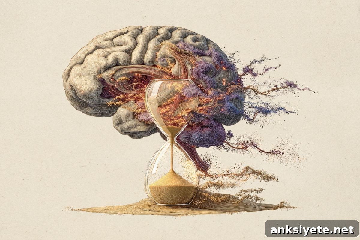
Summary: Scientists have identified how the brain measures time to produce precisely timed actions. Their experiments reveal a neural “hourglass” mechanism in which the motor cortex sends timing signals that accumulate in the striatum. When accumulated activity reaches a threshold, an action is initiated.
Brief interruptions of either region interrupt this internal timer: suppressing the motor cortex temporarily pauses timing, while suppressing the striatum resets it. These results clarify how the brain coordinates movement and suggest avenues for treating timing-related motor disorders such as Parkinson’s and Huntington’s disease.
Key Facts
- Neural hourglass: Timing signals travel from the motor cortex to the striatum and accumulate over time.
- Pause versus reset: Temporarily silencing the motor cortex halts accumulation; silencing the striatum clears the accumulated signal and restarts timing.
- Clinical relevance: Understanding this timing circuit may help design interventions for motor-control disorders that impair movement timing.
Source: Max Planck Institute
Precise, adaptable timing underlies everyday actions, from conversation to sports.
Although we lack sensory organs that detect time directly, the brain keeps internal time to coordinate actions. The specific neural mechanism for that timing function has been unclear. New research from the Max Planck Florida Institute (MPFI) explains how two brain areas—the motor cortex and the striatum—work together to form an adjustable neural timer.
Published in Nature, the study by Zidan Yang, Hidehiko Inagaki, and colleagues shows these regions operate like the top and bottom chambers of an hourglass: the motor cortex sends ramping signals and the striatum integrates them until a movement is triggered.
Discovering the brain’s hourglass
Previous studies pointed to both the motor cortex and the striatum as important for timing. Each area displays ramping neural activity before voluntary movement, and damage to either region produces timing problems in disorders such as Parkinson’s and Huntington’s disease. Still, it remained unclear what unique contribution each region makes to timing and how they interact.
To answer these questions, the researchers trained mice on a simple timing task: a lick for a treat after a set interval (for example, one second). While animals performed the task, the team recorded activity from thousands of neurons across both brain regions to capture their timing-related dynamics.
They combined these recordings with brief, targeted perturbations using optogenetic suppression to transiently silence activity in either the motor cortex or the striatum. By observing how neural signals and behavior changed after each perturbation, the team disentangled the specific roles of each area in the timing process.
Pausing and rewinding the internal timer
The experiments revealed distinct effects. Suppressing the motor cortex interrupted incoming timing signals to the striatum and halted the accumulation of activity there. Behaviorally, this produced a delay roughly equal to the suppression duration—like pinching the neck of an hourglass and pausing the flow of sand.
In contrast, silencing the striatum reduced the accumulated activity and effectively reset the timer. After striatal inhibition, neural activity resumed from a lower baseline and the animal’s response was delayed beyond the suppression period—akin to flipping the hourglass and restarting the count.
These results support a model in which the motor cortex supplies time-varying input and the striatum integrates that input to produce ramping activity that determines when a movement should occur. The motor cortex behaves as the signal source, while the striatum functions as an integrator that sets the timing threshold for action initiation.
Implications for movement and medicine
This work advances our understanding of how distributed neural circuits implement flexible motor timing. By pinpointing how cortical signals are accumulated and converted into timed actions, the study suggests concrete mechanisms that could be targeted to correct timing deficits in movement disorders.
Lead and senior authors emphasize the translational potential: because the motor cortex and striatum are central to movement control and are affected in many motor disorders, mapping their timing roles could guide strategies to restore smoother, more precise movements for affected individuals.
Key Questions Answered:
A: It uses coordinated activity between the motor cortex and the striatum, where cortical signals are integrated in the striatum like sand accumulating in an hourglass.
A: Briefly silencing the motor cortex pauses the timer; briefly silencing the striatum resets it.
A: Because impaired timing in these regions contributes to motor disorders, understanding the circuit could inform new therapeutic approaches.
Editorial Notes:
- This article was edited by a Neuroscience News editor.
- The original journal paper was reviewed in full by staff.
- Additional context was added by editorial staff to clarify implications and methods.
About this movement and neuroscience research news
Author: Lesley Colgan
Source: Max Planck Institute
Contact: Lesley Colgan – Max Planck Institute
Image: The image is credited to Neuroscience News
Original Research: Open access. “Integrator dynamics in the cortico-basal ganglia loop for flexible motor timing” by Zidan Yang et al., published in Nature.
Abstract
Integrator dynamics in the cortico-basal ganglia loop for flexible motor timing
Flexible control of motor timing is essential for behavior. Before voluntary movement begins, frontal cortex and striatum show ramping spiking activity whose slope varies with the expected movement onset. These dynamics across the cortico-basal ganglia loop may serve as an adjustable timer to trigger actions at desired times.
Because frontal cortex and striatum both display similar ramping activity and are necessary for timing behavior, teasing apart their unique contributions has been difficult. To address this, the researchers combined perturbation experiments with multi-regional electrophysiology in mice performing a flexible lick-timing task.
Transient silencing of the frontal cortex produced a rapid recovery of cortical and striatal activity that resumed ramping from pre-silencing levels, shifting lick timing by approximately the duration of the suppression. Brief inhibition of the striatum produced a gradual reduction in ramping across both regions and led to activity resuming from a reduced baseline, shifting lick timing beyond the inhibition period. Thus, inhibiting the frontal cortex and the striatum effectively paused and rewound the timing process, respectively.
These results support a model in which the striatum integrates cortical input to produce ramping activity that controls motor timing, with the motor cortex providing the time-varying drive and the striatum acting as the temporal integrator.