
Summary:
Researchers at the University of California, Berkeley used human stem-cell-derived brain organoids grown for up to a year to model newborn brain development and investigate the origins of tuberous sclerosis complex (TSC) lesions. Their work shows that astrocytes carrying TSC2 mutations adopt an inherently hyperreactive, pro-inflammatory state as they emerge, and these reactive glial cells are central drivers of the lesion formation that triggers severe, drug-resistant pediatric epilepsy. The findings shift the long-standing view that glial inflammation is only a downstream consequence of seizures and suggest that therapies aimed at calming reactive astrocytes could offer focused alternatives to systemic mTOR inhibition.
Key Facts:
- Astrocytes as Primary Drivers: Astrocytes with TSC2 loss arise in a reactive, inflammatory state and directly contribute to tuber development rather than simply responding to seizure activity.
- Long-Term Organoid Culturing: Because human astrocytes mature only around the perinatal or early postnatal period, investigators maintained three-dimensional brain organoids for nine months to a year to capture the transition of radial progenitors into glia.
- Molecular Overlap with Neurodegeneration: Single-cell gene expression analysis revealed that reactive astrocytes in TSC overexpress inflammatory and neurodegeneration-associated genes—an expression pattern confirmed in resected tuber tissue from pediatric patients.
Source: University of California – Berkeley
Rethinking the Origins of Pediatric Seizures
Tuberous sclerosis complex (TSC) is a genetic disorder that affects roughly 1 in 6,000 to 10,000 births and produces cortical malformations, or tubers, that commonly cause early-onset, treatment-resistant epilepsy. Historically, clinicians and researchers have assumed that mutant neurons drive hyperexcitability and that glial inflammation and reactive cells appear later as a consequence of repeated seizures.
The UC Berkeley study, published in Nature, challenges this neuron-centric model. By tracking cell development in long-lived human brain organoids, the team found that astrocytes with TSC2 mutations are born in a disease-like, inflammatory state and actively promote lesion formation. According to senior author Helen Bateup, the astrocytes “look like they’ve been triggered into a disease state” from the moment they arise, indicating a primary role for glia in TSC pathogenesis.
Recreating the Newborn Brain in a Dish
TSC follows a two-hit mechanism: inheritance of one altered copy of TSC1 or TSC2 followed by a somatic second-hit mutation that inactivates the remaining copy, unleashing hyperactive mTOR signaling, a central regulator of cell growth and metabolism. Animal models have provided insights but often fail to recapitulate human-specific developmental timing and pathology.
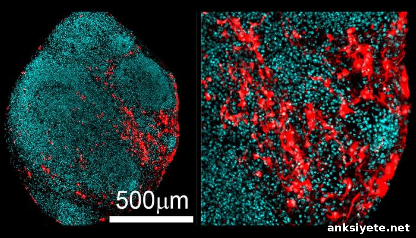
To overcome these limits, the researchers used stem-cell-derived 3D brain organoids and grew them in nutrient-rich culture for extended periods. Human radial glial progenitors produce large numbers of neurons before switching to glial production around birth; capturing that switch required months-long culture. In organoids engineered with TSC2 loss, progenitors diverted prematurely into enlarged, inflammatory astrocytes instead of making healthy neurons, revealing a developmental bias toward reactive glia.
Shared Signatures with Neurodegeneration
Single-cell transcriptomics mapped the gene-expression landscape of individual cells in these organoids. The reactive astrocytes overexpressed inflammatory cytokines, downregulated glutamate transporters, and showed increased expression of genes implicated in neurodegenerative risk—patterns reminiscent of astrocyte states reported in disorders such as Alzheimer’s disease.
Importantly, the team validated these organoid-based signatures in surgical samples: tuber tissue resected from ten pediatric TSC patients exhibited the same abnormal protein and gene-expression profiles, strengthening the link between cell-autonomous glial reactivity and human disease.
Toward Targeted Glial Therapies
Current treatments for mTOR-driven epilepsies (mTORopathies) often rely on systemic mTOR inhibitors like rapalogs, which can reduce seizure frequency but produce broad metabolic and immune side effects because mTOR regulates many essential processes across the body.
By identifying reactive astrocytes as a primary disease driver in TSC, the study opens a path for more selective interventions. Rather than globally suppressing mTOR, clinicians could explore repurposing anti-inflammatory or immunomodulatory drugs to specifically dampen harmful astrocyte activity, restore glial homeostasis, or block astrocyte-mediated damage to surrounding neurons. As Bateup notes, if glia are a core source of pathology, targeted suppression of their inflammatory signaling could potentially reduce seizures without the systemic costs of current therapies.
Funding: The research was supported by the National Institute of Neurological Disorders and Stroke (R01NS097823), a Siebel Stem Cell Center Seed grant, and a Chan Zuckerberg Biohub investigator award. Thomas Li and John Blair are co-first authors. Additional authors include project scientist Taesun Yoo; Professor Dirk Hockemeyer; neurosurgeon Gerald Grant; pediatric neurologist Brenda Porter; and senior author Helen S. Bateup.
Editorial Notes:
- This article was edited by a Neuroscience News editor.
- The journal paper was reviewed in full.
- Additional contextual information was added by staff editors.
About this Epilepsy Research:
- Media Contact: Robert Sanders
- Source: University of California – Berkeley
- Image Credit: Image credited to Helen Bateup/UC Berkeley
- Original Research (Open Access): Nature Communications, September 23, 2026. Title: “mTORC1 drives cell-autonomous astrocyte reactivity in Tuberous Sclerosis.” Authors: Thomas L. Li, John D. Blair, Taesun Yoo, Gerald A. Grant, Dirk Hockemeyer, Brenda E. Porter & Helen S. Bateup.
- DOI: 10.1038/s41586-026-11054-w
Abstract
mTORC1 drives cell-autonomous astrocyte reactivity in Tuberous Sclerosis
Tuberous sclerosis complex (TSC) is a genetic neurodevelopmental disorder marked by focal cortical malformations called tubers, which are frequently associated with severe, intractable epilepsy. Tubers are believed to arise from somatic second-hit mutations that inactivate TSC1 or TSC2 in neural progenitors, leading to hyperactive mTORC1 signaling.
Glial abnormalities are commonly observed in tubers, but it has been unclear whether glia actively drive disease or simply reflect chronic seizure effects. To disentangle these possibilities, the study used human brain organoids to follow mutated progenitors through development in the absence of seizure activity.
Using single-cell transcriptomics and cyclic immunostaining across organoids and resected human tuber tissue, the researchers show that loss of TSC2 biases progenitors to form enlarged, pro-inflammatory astrocytes in a cell-autonomous manner. These mutant astrocytes exhibit reduced glutamate transporter levels, elevated inflammatory cytokine secretion, and increased expression of neurodegeneration-associated genes such as APOE and CLU.
The findings demonstrate that reactive astrocytes emerge as a primary consequence of TSC2 loss, implicating glial dysfunction as a driver of TSC pathogenesis and highlighting reactive astrocytes as promising therapeutic targets for TSC-related neuropathology.