
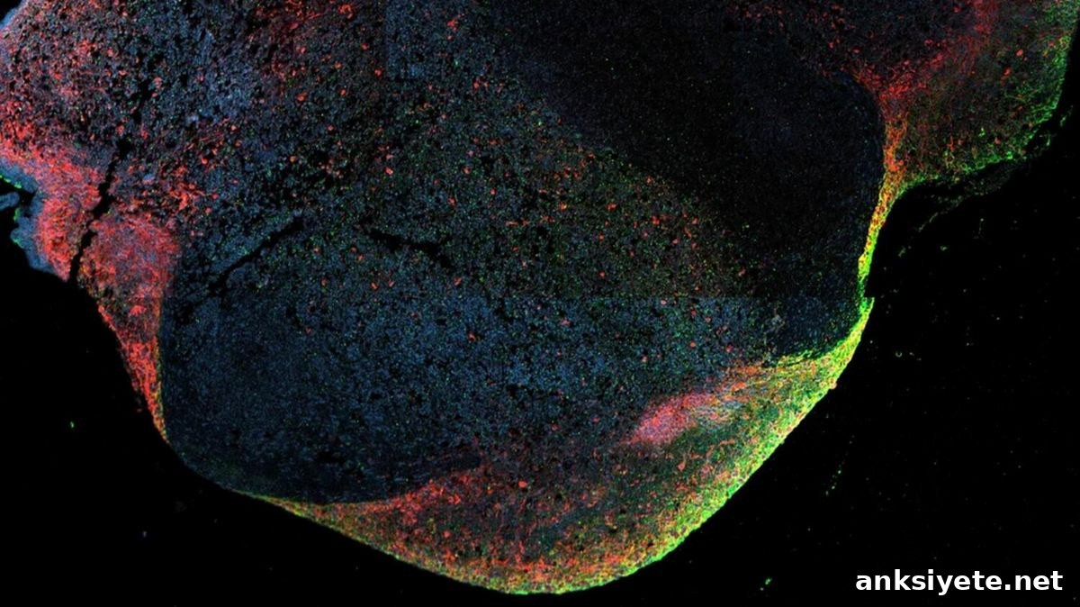
Summary: Researchers have uncovered how the Ebola virus can persist undetected in the human central nervous system for months or even years. An international team used human induced pluripotent stem cells (iPSCs) to grow three-dimensional cerebral organoids and studied long-term infections in a fully human tissue model.
The study shows that Ebola and related filoviruses (including Sudan, Reston, and Marburg viruses) establish a form of “productive persistence” in brain tissue. Rather than remaining completely inactive, the virus continues to replicate inside neurons, astrocytes, and microglia for up to 120 days. During this time it generates defective viral genomes and triggers chronic, localized inflammation that mirrors the severe meningoencephalitis observed in some human survivors.
Key Facts
- Productive persistence, not latency: Contrary to the assumption that long-term viral survival requires complete dormancy, this work demonstrates that Ebola can persist by ongoing replication, producing infectious particles over extended periods without immediately destroying host tissue.
- Two transmission modes: Within brain organoids the virus spreads both by classical budding—releasing free virions from the cell membrane—and by direct cell-to-cell transmission, moving between adjacent neurons and thereby avoiding exposure in the extracellular space.
- Chronic, local inflammation: Although the brain is an immune-privileged site that limits systemic immune responses to protect delicate tissue, persistently infected organoids developed a late surge of pro-inflammatory cytokines. This pattern is consistent with the eye, meningeal, and brain inflammation seen months after acute Ebola infection in survivors.
- Defective genomes as a survival strategy: Researchers detected defective viral genomes and specific mutations that appear to attenuate replication. Because filoviruses lack high-fidelity proofreading, replication errors accumulate; some of these altered genomes reduce replication speed and help maintain a long-term, sublethal infection.
- Human-relevant mutations: Many of the mutations seen in organoids matched variants previously isolated from human outbreaks. This concordance supports the organoid model as a translationally relevant system for studying human-specific viral adaptation.
- Reducing reliance on animal models: Conducting prolonged infections in a 100% human genetic background offers a practical platform for screening antivirals and studying pathogenesis while minimizing dependence on high-containment animal experiments.
Source: DZIF
After recovery from acute disease, Ebola virus can remain hidden in immune‑privileged sites such as the central nervous system for months or even years, creating the potential for relapse or—rarely—renewed transmission.
Researchers from the Bernhard Nocht Institute for Tropical Medicine (BNITM), the German Center for Infection Research (DZIF), the Icahn School of Medicine at Mount Sinai (ISMMS), and collaborators used a cerebral organoid model to investigate how Ebola persists in the brain. Their findings were published in Nature Microbiology.
Ebola virus disease is a severe multisystem infection with a high case-fatality rate. Even after surviving the acute phase, infectious virus can persist in body compartments with limited immune surveillance—semen is one well-documented example, and the central nervous system is another. Because immune activity is dampened in these sites to protect sensitive structures, immune clearance can be incomplete and persistent virus may remain a long-term health risk.
Cerebral organoids as a model for Ebola persistence
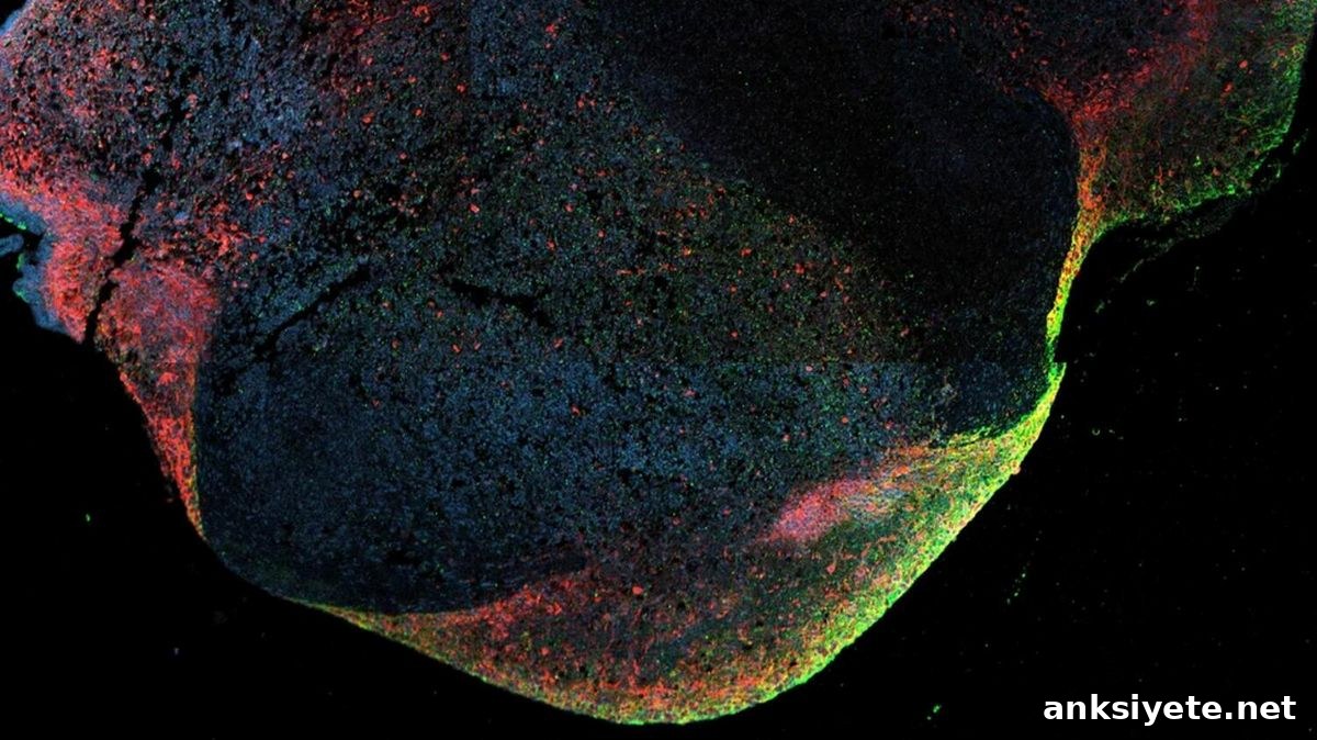
Studying persistence in the human central nervous system is challenging. To overcome this, the team used established cerebral organoids derived from human iPSCs. These spherical, brain-like tissues contain multiple central nervous system cell types—neurons, astrocytes, and microglia—and provide a scalable human platform to study host–virus interactions over extended periods.
“Cerebral organoids enable detailed investigation of how Ebola and other filoviruses persist in the human CNS,” says Dr. Lina Widerspick, first author and former BNITM researcher. “This model helps explain the mechanisms behind the severe and sometimes fatal inflammation seen in survivors with meningoencephalitis.”
Organoids also offer a human-relevant alternative to animal models, improving translational relevance for antiviral testing and reducing reliance on animal experiments in high-containment facilities.
Sustained replication and cell tropism in organoids
The investigators found that Ebola and related filoviruses replicated in cerebral organoids for up to 120 days. Multiple cell types were infected: neurons and astrocytes supported viral replication, and microglia—the brain’s resident immune cells—were recruited to infection sites and became infected themselves.
Viral spread occurred through both cell-to-cell transmission and budding of free virions, demonstrating productive persistence: the virus remained infectious rather than entering a purely dormant state. Although organoids mounted innate immune responses and produced inflammatory cytokines, these responses did not clear the infection during the study period.
“We saw elevated immune and inflammatory signals late in organoid cultures,” notes Prof. César Muñoz-Fontela of BNITM. “This supports the idea that persistent Ebola infection in immune‑privileged tissues can drive local inflammation, consistent with clinical observations in some survivors.”
Viral adaptation during persistence
Defective viral genomes are a common viral strategy to limit replication and support long-term survival in the host. The team identified defective genomes and specific mutations accumulating in late-stage infected organoids. Many of these variants correspond to mutations previously suggested to reduce replication in patients, validating the organoid model’s ability to recapitulate human viral evolution.
Some mutations discovered in organoids have not yet been documented in survivors; further work is required to determine whether those changes contribute causally to persistence.
“Because Ebola behaves in this human model much like it does in infections, cerebral organoids are a powerful tool to study filovirus persistence and to test therapeutic strategies,” adds Prof. Gustavo Palacios of ISMMS.
The authors emphasize the need for continued research into long-term virus–host interactions, including studies of less-characterized filoviruses such as Reston, Taï Forest, Bombali, and Bundibugyo, to deepen understanding of persistence mechanisms.
Funding: In addition to BNITM, ISMMS, and IRF‑Frederick/NIH, collaborators included University Medical Center Hamburg‑Eppendorf (UKE), Leibniz Institute of Virology (LIV), and the Friedrich‑Loeffler‑Institut (FLI). Financial support came from sources including the Collaborative Research Center 1648 “Emerging Viruses” of the German Research Foundation (DFG), the German Federal Institute for Risk Assessment (BfR), and DZIF.
Key Questions Answered:
A: The virus establishes productive persistence in immune‑privileged regions where immune responses are naturally limited. By producing defective genomes and attenuated variants, Ebola slows its replication so it does not rapidly destroy host tissue, allowing a prolonged, low‑level infection that escapes complete immune clearance.
A: A cerebral organoid is a miniature, three‑dimensional human brain model grown from stem cells. It contains human neurons, astrocytes, and immune cells in organized structures, making it a human‑relevant platform to observe viral behavior and host responses that are difficult to replicate in animal models.
A: The study helps explain persistent neurological symptoms some survivors experience—such as vision problems, headaches, and meningoencephalitis—by showing that active viral replication in the CNS can drive local inflammation. The results underscore the importance of long‑term medical follow‑up and targeted antiviral strategies for survivors.
Editorial Notes:
- This article was edited by a Neuroscience News editor.
- The original journal paper was reviewed in full.
- Additional explanatory context was added by staff to clarify findings and implications.
About this neurology and Ebola research news
Author: Press Office
Source: DZIF
Contact: Press Office – DZIF
Image: The image is credited to BNITM/Lina Widerspick
Original Research: Open access. “Host–virus determinants of Ebola virus persistence in a human cerebral organoid model” by Lina Widerspick et al., Nature Microbiology. DOI: 10.1038/s41564-026-02388-2
Abstract
Host–virus determinants of Ebola virus persistence in a human cerebral organoid model
Ebola virus (EBOV) causes Ebola virus disease (EVD), a multisystem human illness with a high fatality rate. Survivors may develop recurrent inflammation linked to viral persistence in immune‑privileged tissues, including the central nervous system. Persistence—defined as ongoing EBOV genome replication beyond the acute phase—can produce virions and has been implicated in re‑initiating outbreaks. Using a human cerebral organoid model, the authors show EBOV persistence for 120 days, sustained by continuous infection of astrocytes and neurons, recruitment and infection of microglia, emergence of defective genomes and subvariants, cell‑to‑cell transmission, activation of cell‑specific innate immunity, and late organoid inflammation, indicating that persistent EBOV infection within immune‑privileged niches drives local inflammation.