
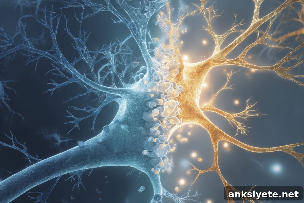
Summary: New research shows that astrocytes in the lateral hypothalamus are central to how stress — especially stress experienced early in life — reshapes behavior. In mice, early-life stress produced smaller, less branched astrocytes and altered activity of orexin-producing neurons, with opposite activity patterns emerging in males and females.
When the research team removed stress hormone receptors specifically from astrocytes, they were able to restore normal neuronal firing and behavioral patterns, even though astrocyte size did not fully revert to its original state. These results point to astrocytes as promising targets for interventions aimed at preventing stress-linked, treatment-resistant depression.
Key Facts
- Astrocyte-first response: Stress alters astrocyte structure and function before neurons show measurable dysfunction.
- Sex-specific outcomes: Early-life stress produced opposite changes in activity between male and female mice.
- Therapeutic insight: Deleting glucocorticoid receptors in lateral hypothalamic astrocytes normalized neuron firing and behavior.
Source: University of Montreal
Astrocytes in the lateral hypothalamus — a brain region involved in sleep-wake regulation — strongly influence neuronal firing and behavior in mice, according to Canadian researchers.
Led by Ciaran Murphy-Royal at Université de Montréal’s affiliated hospital research centre (CRCHUM), the team reports their findings in Nature Communications. Their work expands our understanding of brain circuitry and highlights how non-neuronal cells can drive long-term changes in behavior after early stress.
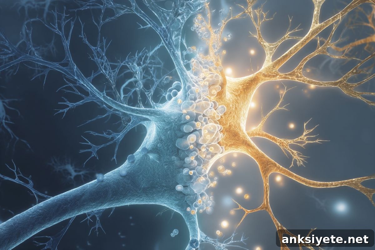
The researchers emphasize that these discoveries could eventually inform treatments to prevent or reduce the impact of depression that arises after adverse childhood experiences. Prior studies already link early-life stress to a markedly elevated risk of adult mental-health disorders, including forms of depression that resist conventional therapies.
Behavioral changes and stress hormones
Astrocytes respond to changes in blood-borne metabolites and hormones by remodeling their shape and altering how they interact with nearby neurons. In rodents, corticosterone is the primary stress hormone, and the study found chronically elevated corticosterone levels in adult mice that had been exposed to stress in early life.
Murphy-Royal notes a clear sex difference in the behavioral consequences: females showed reduced nighttime activity, while males displayed increased daytime activity. Similar sex-specific activity patterns have been documented in humans who experienced comparable early-life stress, particularly among individuals with depressive disorders.
Modeling lack of maternal care
To model early-life adversity, first author Lewis R. Depaauw-Holt separated mouse pups from their mothers for four hours daily over a ten-day window that corresponds to a critical developmental period — roughly analogous to ages three to seven in human children. This manipulation produced long-lasting changes in activity and brain physiology.
The team focused on orexin-producing neurons in the lateral hypothalamus, cells known to regulate arousal, motivation, and sleep–wake cycles. In stressed animals, orexin neurons were hyperactive in males but hypoactive in females, mirroring the opposing behavioral phenotypes.
Concurrently, astrocytes in the lateral hypothalamus became smaller and lost branching complexity, with females showing the most pronounced morphological reductions. Because astrocytic processes mediate communication with neurons and other supporting cells, their retraction is interpreted as a sign of dysfunction that can disrupt local neural circuits.
One stress-signaling pathway
To determine whether a single molecular pathway might link stress, astrocyte changes, and altered neuronal activity, the researchers selectively deleted glucocorticoid receptors from astrocytes in the lateral hypothalamus. Glucocorticoid receptors are the principal binding sites for corticosterone.
Remarkably, removing these receptors restored orexin neuron firing rates and normalized activity patterns in both sexes, despite astrocytes not fully regaining their original size. Instead, astrocytes recovered structural complexity—the branching essential for interactions with neighboring cells—suggesting functional recovery even if cell volume stayed reduced.
The study further identified distinct astrocyte-mediated mechanisms driving the sex differences: increased purinergic signaling appeared to underlie male hyperactivity, while reduced extracellular L-lactate was associated with female hypoactivity. These divergent pathways converge on the same glial cell population but produce opposite effects on neuronal output.
Contrary to prevailing assumptions, the data indicate astrocytes are altered by stress before measurable neuronal changes occur, positioning them as primary drivers of long-term behavioral shifts following early adversity.
Murphy-Royal cautions that translating these findings to humans will be complex, but he underscores the therapeutic promise: targeting astrocyte signaling pathways could offer new strategies to prevent or treat stress-related psychiatric conditions, including forms of depression resistant to current treatments.
About this neuroscience and mental health research news
Author: Bruno Geoffroy
Source: University of Montreal
Contact: Bruno Geoffroy – University of Montreal
Image: Image credit: Neuroscience News
Original Research: Open access. “A divergent astrocytic response to stress alters activity patterns via distinct mechanisms in male and female mice” by Ciaran Murphy-Royal et al., Nature Communications.
Abstract
A divergent astrocytic response to stress alters activity patterns via distinct mechanisms in male and female mice
The lateral hypothalamus regulates overall activity, circadian rhythms, and motivated behaviour. Although disruption of these functions is central to many stress-related psychiatric disorders, the cellular mechanisms by which stress acts on this region are not well understood.
This study shows that stress-driven alterations in spontaneous orexin neuron firing correspond with divergent activity patterns: increased activity in males and decreased activity in females. These neuronal changes coincide with remodeling of astrocytes; experiments demonstrate that lateral hypothalamic astrocytes causally regulate orexin neuron firing and whole-animal activity.
Under stress, sex-specific astrocytic mechanisms emerge: elevated purinergic signaling in males and diminished extracellular L-lactate in females drive the distinct neuronal responses. Genetic removal of glucocorticoid receptors from lateral hypothalamic astrocytes restores key aspects of astrocyte morphology, normalizes orexin firing, and recovers activity levels in both sexes.
Together, these results establish astrocytes as primary mediators of stress-induced behavioral changes and key regulators of orexin neuron activity and physical activity patterns.