
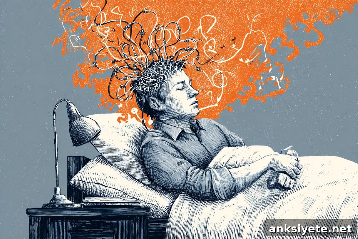
Summary: Researchers have reproduced the restorative effects of sleep in small, targeted areas of the brain in awake mice. Using optogenetic stimulation, they generated a rhythmic, alternating “on-and-off” firing pattern — the defining signature of non-rapid eye movement (NREM) slow-wave sleep — for 30-minute periods. The intervention reversed memory impairments caused by sleep deprivation and reduced the local biological need for sleep in the stimulated regions, demonstrating that sleep’s restorative power depends on specific slow-wave rhythms rather than a mere global decrease in neuronal firing.
Key Facts
- Local induction of sleep-like activity: The team successfully evoked sleep-like neural patterns in confined regions of the cortex while the rest of the brain remained awake, alert, and responsive to the environment.
- Genetic and light-based method: The experiment combined genetic targeting with implantable light-delivery devices to impose rhythmic on/off patterns in selected cortical areas for half-hour sessions.
- NREM’s selective pruning role: During NREM sleep, the brain evaluates synaptic connections—protecting essential links for long-term memory while pruning weaker ones to free capacity for new learning.
- Against a simple “neuronal fatigue” model: Results show that recovery requires the specific alternating slow-wave pattern of activity, not just an overall reduction in neuronal firing that some have proposed as recovery from wake-induced fatigue.
- Reduced local sleep pressure: After stimulation, those same cortical regions showed lower slow-wave activity during subsequent sleep, indicating that their local sleep need had been met.
- Memory preservation: In tactile memory tests dependent on sleep, sleep-deprived mice that received local stimulation performed as well as rested controls, while deprived mice without stimulation performed worse.
- Human translation prospects: Corresponding author Chiara Cirelli plans to investigate whether noninvasive transcranial stimulation in humans can reproduce these localized restorative patterns to help protect cognition.
Source: NIH
By inducing precise on/off activity in small cortical regions of awake mice, researchers supported by the National Institutes of Health (NIH) triggered synaptic recalibration normally reserved for sleep. This localized approach counteracted the effects of sleep deprivation on memory and clarified mechanisms central to sleep’s restorative function.
“What we’re essentially doing is forcing sleep in a local region of the brain. While that part is solidifying memories and restoring learning capacity, other parts stay aware and connected to the environment,” said corresponding author Chiara Cirelli, M.D., Ph.D., professor of psychiatry at the University of Wisconsin–Madison. “Some animals, like dolphins, naturally exhibit one-hemisphere sleep, which illustrates how sleep can be regional.”
NREM sleep, which constitutes roughly 80% of adult sleep time, is when neuronal junctions involved in memory are evaluated. During NREM, the brain preserves important synapses for long-term storage, removes less useful connections, and makes room for future learning.
Prior work from Cirelli and colleagues found that rats and humans can show local slow-wave activity while awake after sleep deprivation, but those spontaneous episodes were brief and inconsistent, leaving open whether prolonged, patterned induction could reproduce sleep’s benefits. The current study tested that idea directly.
Using optogenetics, the researchers induced alternating on/off periods in targeted cortical zones of sleep-deprived mice for 30-minute sessions, imitating the temporal structure of slow waves during NREM sleep. When the mice later slept, slow-wave activity and synchrony were reduced specifically in the stimulated areas, indicating that those regions had lower residual sleep pressure.
Further experiments showed that the beneficial effects depended on the rhythmic alternation itself. Simply lowering overall firing did not produce the same outcome, countering the hypothesis that recovery from wakefulness is achieved merely by a generalized neuronal quieting.
To test behavioral consequences, the team measured tactile memory, a sleep-sensitive cognitive function. Sleep-deprived mice that received bilateral stimulation across motor and sensory regions performed on par with well-rested controls, while deprived mice without stimulation showed clear deficits.
Cirelli and collaborators plan to explore whether noninvasive transcranial approaches can elicit comparable localized slow-wave patterns in humans, with the long-term goal of mitigating cognitive decline and improving memory resilience.
“This research helps decode why we sleep and how learning is consolidated,” said Amy Bany Adams, Ph.D., acting director of NIH’s National Institute of Neurological Disorders and Stroke (NINDS), which funded the work. “Understanding these mechanisms brings us closer to strategies for preventing and treating cognitive impairment.”
Key Questions Answered:
A: The researchers targeted a small cortical area with optogenetic tools and light pulses to force a localized, rhythmic on/off firing pattern that mirrors NREM slow waves. This created sleep-like physiology in that region while surrounding brain areas remained awake and behaviorally responsive.
A: No. The study indicates that recovery requires the specific slow-wave timing — alternating active and silent periods — rather than a simple overall decrease in firing rates.
A: It is an aspirational goal. The current findings were obtained with invasive implants in mice. Researchers are now exploring whether noninvasive transcranial stimulation can reproduce the same local slow-wave patterns in humans, but clinical application will require further study and validation.
Editorial Notes:
- This article was edited by a Neuroscience News editor.
- Journal paper reviewed in full.
- Additional context provided by the editorial staff.
About this sleep and brain stimulation news
Author: Jonathan Griffin
Source: NIH
Contact: Jonathan Griffin – NIH
Image: The image is credited to Neuroscience News
Original Research: Open access. “Induction of cortical ON/OFF periods in awake mice fulfills sleep functions” by Kort Driessen, Fabio Squarcio, Giulio Tononi & Chiara Cirelli. Nature Neuroscience. DOI: 10.1038/s41593-026-02318-9
Abstract
Induction of cortical ON/OFF periods in awake mice fulfills sleep functions
Slow-wave sleep in mammals is defined by synchronized neuronal activity that alternates between active (ON) and silent (OFF) periods. Slow-wave activity (SWA) and neuronal synchrony are correlated with sleep need, reflect synaptic strength in cortical circuits, and support synaptic downselection and memory consolidation. This study asked whether those core sleep benefits can be produced during wakefulness.
Using optogenetics, alternating ON/OFF periods were locally induced in awake mice. This manipulation reduced ipsilateral SWA and synchrony during the mice’s subsequent sleep and lowered markers of synaptic strength in the stimulated regions. Bilateral induction of OFF periods over the sensorimotor cortex during sleep deprivation restored memory consolidation.
These results show that driving ON/OFF activity during wakefulness is sufficient to reduce local sleep need and to fulfill essential functions typically achieved during sleep.