
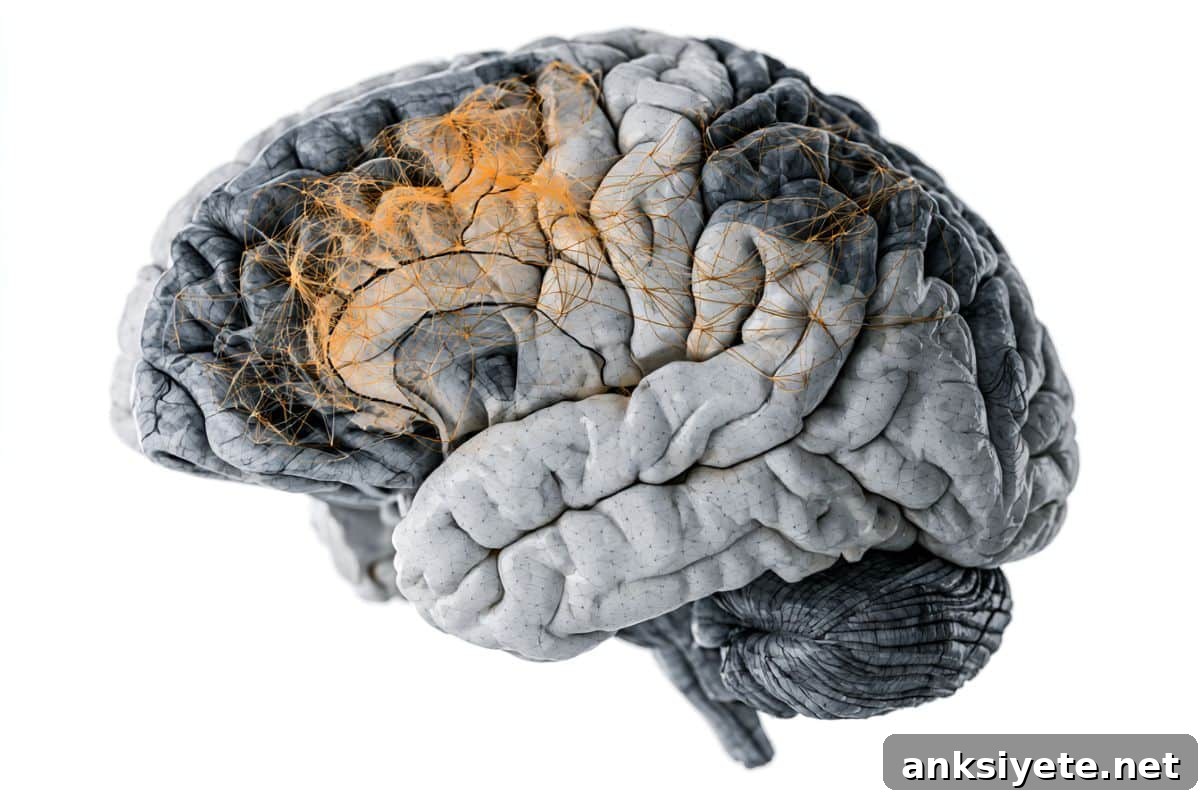
Summary: A new study used specialized positron emission tomography (PET) to measure synaptic connections in living human brains. Analyzing one of the largest clinical cohorts to date, researchers found a clear, widespread reduction in synaptic density in schizophrenia that follows the brain’s underlying molecular and network architecture. Their results map a distinct structural starting point and reveal how synaptic breakdown may spread through neural circuits.
Key Facts
- Direct measurement of synapses: Rather than relying on standard anatomical scans, the study used [11C]UCB-J PET imaging to quantify microscopic synaptic connections in living people, providing a direct in vivo measure of synaptic density.
- Widespread synaptic loss: Individuals diagnosed with schizophrenia showed a pronounced, system-wide reduction in synaptic density, most prominently across frontal, temporal, limbic, and memory-related brain regions.
- Left hemisphere vulnerability: Synaptic loss was markedly asymmetric, with the left hemisphere showing substantially greater reduction in synaptic density than the right.
- PET and MRI capture different processes: The spatial pattern of synaptic loss detected by PET did not align with grey matter volume changes measured by standard MRI, indicating these are distinct biological processes.
- Molecularly guided vulnerability: Areas with the greatest synaptic decline corresponded to regions naturally rich in receptors for serotonin, glutamate, and GABA, suggesting regional molecular signatures shape vulnerability to schizophrenia-related damage.
- Identified structural epicenter: Network diffusion simulations pointed to a specific locus in the left frontal lobe as a likely origin from which synaptic pathology spreads to connected and molecularly similar regions.
Source: Rutgers University
New evidence from Rutgers and collaborating institutions reveals how schizophrenia affects synaptic connections across the living human brain.
Published in Molecular Psychiatry, the study was led by Avram Holmes (associate professor of psychiatry, Robert Wood Johnson Medical School; Rutgers Brain Health Institute) and Rajiv Radhakrishnan (associate professor of psychiatry and radiology and biomedical imaging, Yale University). The first author, Sidhant Chopra, completed this work while a postdoctoral fellow in the Holmes Lab and is now affiliated with Orygen and the University of Melbourne.
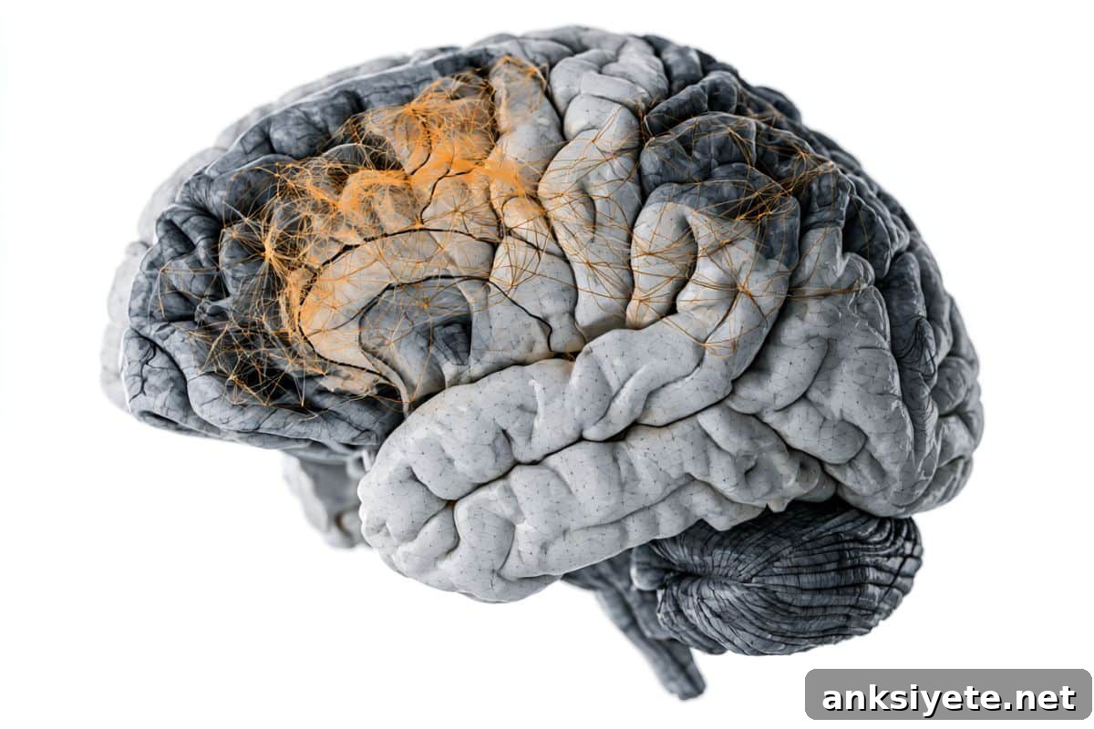
Synapses are the microscopic contact points that allow neurons to communicate across circuits. Disruption of synaptic connections is widely thought to underlie cognitive and emotional symptoms in schizophrenia. Until now, the detailed spatial pattern of synaptic loss in living human brains was difficult to map because conventional magnetic resonance imaging (MRI) cannot directly measure synaptic proteins or their density.
This study analyzed PET scans from 122 participants—29 individuals with schizophrenia and 93 healthy controls—making it one of the largest examinations of synaptic density in psychiatric illness using PET. The researchers found a clear, widespread reduction in synaptic density in people with schizophrenia compared with controls. Losses were especially pronounced in frontal and temporal cortices, cingulate regions, thalamus, striatum, and hippocampus, and were significantly greater in the left hemisphere.
Importantly, the pattern of reduced synaptic density did not overlap with regional grey matter volume changes detectable on MRI. That dissociation suggests synaptic pathology and macroscopic tissue volume loss are separate biological phenomena, each requiring distinct approaches to detection and treatment.
The investigators also examined the relationship between synaptic loss and regional neurochemistry. Regions showing the greatest reduction in synaptic density corresponded to areas that normally have high concentrations of receptors for key neurotransmitters—GABAergic (GABAA/BZ), serotonergic (5HT2A and 5HT1B), and glutamatergic (mGluR5) systems—indicating that a region’s baseline molecular profile predicts its vulnerability in schizophrenia.
Using simulation-based network diffusion models, the team traced likely pathways for how synaptic loss propagates along structural connections. These models identified left inferior frontal regions as the most probable starting locations for pathology, suggesting synaptic damage may originate in a focal left frontal epicenter and then spread to anatomically connected and molecularly similar regions.
“The pattern of synaptic loss in schizophrenia appears organized and predictable,” said Sidhant Chopra. “It follows the brain’s molecular and connectivity architecture, which could guide efforts to prevent, slow, or reverse synaptic decline.”
Avram Holmes added that “this detailed mapping of synaptic vulnerability points to precise targets for emerging therapies aimed at protecting or restoring synapses, offering a path toward more proactive and localized interventions.”
The research team notes future studies should track how synaptic density changes over time, examine the effects of treatments, and refine personalized strategies to preserve neural connectivity and cognitive function in people with schizophrenia.
Key Questions Answered:
A: MRI is excellent at measuring large-scale anatomy and tissue volume but cannot resolve submicroscopic structures like synapses. PET imaging with a tracer that binds synaptic proteins directly measures synaptic density, revealing changes invisible to anatomical MRI.
A: It means that synaptic decline is not random. Regions with high concentrations of receptors for serotonin, glutamate, and GABA show the most loss, implying that a region’s baseline neurochemical profile determines its susceptibility to schizophrenia-related damage.
A: Locating a likely epicenter enables more targeted research and therapy development. Instead of broadly treating symptoms after widespread damage, clinicians and researchers could pursue preventative or localized interventions intended to protect or restore synapses before pathology spreads.
Editorial Notes:
- This article was edited by a Neuroscience News editor.
- The full journal paper was reviewed for accuracy.
- Additional context was added by editorial staff.
About this schizophrenia research news
Author: Patti Zielinski
Source: Rutgers University
Contact: Patti Zielinski – Rutgers University
Image credit: Neuroscience News
Original Research: Open access. “Widespread synaptic density loss in schizophrenia follows molecular and network architecture” by Sidhant Chopra, Patrick D. Worhunsky, Mika Naganawa, Xi-Han Zhang, Ashlea Segal, Loïc Labache, Edwina Orchard, Vanessa Cropley, Stephen Wood, Gustavo A. Angarita, Kelly Cosgrove, David Matuskey, Nabeel B. Nabulsi, Yiyun Huang, Richard E. Carson, Irina Esterlis, Patrick D. Skosnik, Deepak C. D’Souza, Avram J. Holmes & Rajiv Radhakrishnan. Molecular Psychiatry. DOI: 10.1038/s41380-026-03717-x.
Abstract
Widespread synaptic density loss in schizophrenia follows molecular and network architecture
Converging neuroimaging, genetic, and post-mortem evidence highlights the central role of reduced synaptic density in schizophrenia. Yet the full brain-wide spatial pattern and mechanisms driving that pattern have remained unclear. Using [11C]UCB-J PET in people with schizophrenia (n = 29) and healthy controls (n = 93), the study identified a prominent, widespread reduction in synaptic density across multiple regions (0.58 < Cohen’s D < 1.47; pFWE < 0.05). The left hemisphere showed significantly greater impact than the right (Cohen’s D = 1.14; p < 0.001), with frontal, temporal, cingulate, thalamic, striatal and hippocampal areas particularly affected.
Synaptic density changes did not spatially correspond to grey matter volume alterations from anatomical MRI. Lower left-hemisphere synaptic density correlated with higher normative concentrations of GABAA/BZ, 5HT2A, mGluR5 and 5HT1B receptors (rcca = 0.68; p = 0.022). Network diffusion simulations nominated left inferior frontal regions (pFWE < 0.05) as plausible initiation sites from which synaptic pathology propagates along axonal networks to structurally connected and molecularly similar areas. These in vivo findings indicate left-lateralised, widespread synaptic deficits in schizophrenia that are independent of grey matter volume change, aligned with specific neurochemical systems, and consistent with propagation along brain networks.