
Summary: Researchers have created a biohybrid neural implant that can strengthen communication between the brain and paralyzed limbs, offering a promising route to restore limb function after amputation or nerve injury.
Source: University of Cambridge
New biohybrid neural implant developed to restore limb function
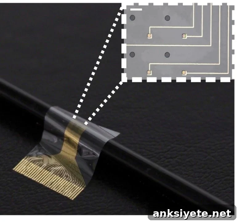
Scientists at the University of Cambridge have developed a biohybrid neural implant that combines flexible electronics with human stem-cell–derived muscle cells to improve the interface between peripheral nerves and prosthetic or reinnervated tissue. In rat experiments, the device enhanced signal capture from nerves that previously failed to communicate effectively with electronic electrodes because of scar tissue and poor integration.
Conventional neural implants often provoke scar formation around electrodes, which degrades signal quality and limits long-term performance. To overcome this, the Cambridge team inserted a thin layer of induced pluripotent stem cell (iPSC)–derived muscle cells between the nerve and a soft, flexible electrode array. The living cell layer acted as an intermediary that the body accepted more readily than bare metal or polymer electrodes, preventing scar encapsulation and improving electrical coupling.
The biohybrid device is designed to sit at the end of a severed or re-routed nerve. Individual axons produce very small voltages, but when an axon connects with a muscle cell the resulting electrical signal is much larger and easier to detect. By using engineered muscle cells as biological amplifiers on the electrode surface, the implant increases sensitivity and enables more detailed readout of motor commands at a near-axon scale.
In freely moving rats, the implanted cell layer survived and integrated with the host nerve throughout a 28-day study—the first time survival of this type of human iPSC-derived muscle cell has been documented over such a period in a living organism. Although the experiments did not restore forearm movement in these animals, the device successfully recorded brain-generated motor signals that would be required to drive a limb or a prosthetic, demonstrating the potential to restore functional control if connected to distal nerve pathways or an actuator.
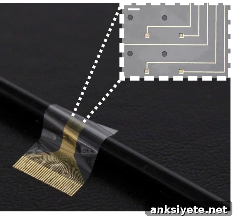
By combining two advanced approaches—cell therapy and bioelectronics—into one compact implant, the researchers addressed limitations that each method faces when used alone. The biological layer prevents foreign-body reactions and acts as a targeted interface that can amplify and spatially resolve neural signals. The flexible electronics provide the signal readout and the mechanical compliance necessary for long-term implantation.
Key advantages of this approach include improved long-term stability, finer resolution of motor signals, and a compact form factor that could be implanted through minimally invasive, keyhole surgery. Unlike some cortical-based prosthetic systems that require complex patient-specific decoding of brain activity, this peripheral nerve interface can scale using standardized, manufactured cell lines and targets the specific axons responsible for motor control.
The implanted muscle cells used in the study were produced using the opti-ox precision reprogramming system, which enables consistent, scalable generation of cell types from iPSCs. The cell lines for this experiment were supplied by the Kotter lab at the University of Cambridge. The opti-ox technology is owned by bit.bio.
Lead researchers emphasize that significant additional development and testing will be required before human clinical use is possible, including extended-duration studies, safety evaluation, and demonstration of restored functional movement in models that reconnect the implant to muscles or prosthetic devices. Nevertheless, the results mark a promising step toward regenerative bioelectronic therapies that could benefit amputees and people with paralysis or peripheral nerve injuries.
Co-first authors reported that integrating living human cells with bioelectronic materials produced a more natural, intuitive communication channel with the nervous system. The team has filed a patent application to protect the platform and is working on device optimization and scale-up.
Funding: This research received partial support from the Engineering and Physical Sciences Research Council (EPSRC) under UK Research and Innovation (UKRI), Wellcome, and the European Union’s Horizon 2020 Research and Innovation Programme.
About this paralysis and neurotechnology research
Author: Sarah Collins
Source: University of Cambridge
Contact: Sarah Collins, University of Cambridge
Image: Image credit: University of Cambridge
Original Research: Open access. “Functional neurological restoration of amputated peripheral nerve using biohybrid regenerative bioelectronics” by Damiano Barone et al., Science Advances. DOI and journal reference available in the original publication.
Abstract
Functional neurological restoration of amputated peripheral nerve using biohybrid regenerative bioelectronics
Development of neural interfaces with improved biocompatibility and tissue integration is essential for treating neurological damage and restoring function. Increasing the resolution for mapping neuronal inputs onto implants is a critical challenge. The authors introduce a new category of neural interface in which iPSC-derived myocytes are grafted onto flexible electrode arrays to serve as biological targets for peripheral nerve inputs. They demonstrate long-term survival and functional integration of human iPSC-derived cells with a forearm nerve bundle in freely moving rats over four weeks, and show that the intermediate cell layer enhances tissue-electronics coupling and electrical recording in vivo, representing a step toward restorative therapies using regenerative bioelectronics.