
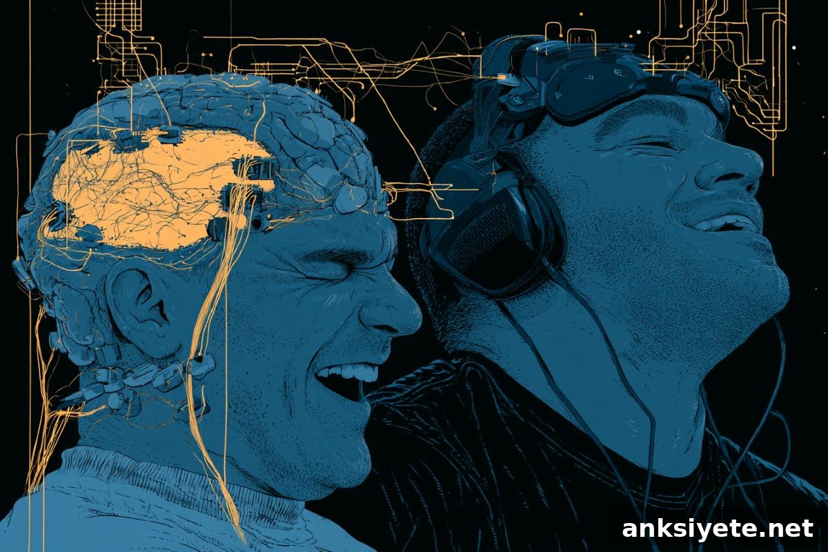
Summary: Laughter is one of humanity’s most universal social signals, but pinpointing the exact brain circuits that produce it has been difficult because genuine, unforced mirth is hard to elicit in laboratory settings. A new review of clinical stimulation data from awake pre-surgical epilepsy patients provides rare causal evidence linking specific brain regions to either spontaneous, emotion-driven laughter or deliberate, conversational laughter.
Researchers compiled and reanalyzed reports from awake brain-mapping procedures in which clinicians temporarily stimulate localized areas of the cortex and subcortex to identify surgical targets. When electrical probes inadvertently triggered involuntary laughter, patients could report whether the experience included real feelings of joy or was purely a motor response. Combining these clinical observations with complementary animal and clinical studies, the authors outline a dual-network model for the neural basis of laughter.
Key Facts
- Two distinct neural systems: Human laughter is supported by two anatomically and functionally separate networks — an older, emotion-driven spontaneous system and a cortical, motor-based volitional system.
- Clinical mapping in awake patients: Data come largely from pre-surgical cortical and subcortical stimulation performed while epilepsy patients are awake. Those rare, real-time reports let researchers distinguish authentic mirth from simple facial or respiratory motor patterns.
- Spontaneous network evokes mirth: Stimulating regions such as the pregenual anterior cingulate cortex, nucleus accumbens, and temporal pole provokes intense, genuine laughter often accompanied by euphoria and a clear change in mood.
- Volitional network controls the mechanics: Stimulation of motor-related areas — including the rolandic operculum, globus pallidus, and presupplementary motor area — produces laughing and smiling movements without any accompanying positive emotion.
- Evolutionary perspective: The spontaneous system appears evolutionarily ancient and likely evolved from play-related vocalizations (rough-and-tumble signals) that reduce aggression and strengthen social bonds across mammals.
- Analgesic links: The anterior cingulate’s role within the spontaneous network ties laughter to the brain’s pain-modulation systems, offering a plausible mechanism for why laughter can temporarily raise pain thresholds.
Source: Cell Press
Why this matters: Laughter is a powerful social cue that binds people together, but the brain circuits that generate it have been poorly defined. The new synthesis highlights how different neural pathways produce either involuntary, emotion-laden laughter or precisely timed, socially used laughter. This distinction clarifies longstanding observations from clinical neurology and everyday social behavior.
The review, published June 23 in the journal Trends in Neurosciences, integrates direct human stimulation reports with other clinical and animal research. The authors emphasize two qualitatively different kinds of laughter people commonly experience. The first — spontaneous laughter — is an involuntary, often overwhelming reaction: “something sets you off and you are helpless with mirth,” as the authors note. This form is sometimes observed in certain neurological or psychiatric conditions, including some seizure disorders and neurodegenerative illnesses.
The second form is volitional laughter: the deliberate, tightly timed laughs that punctuate conversation. Volitional laughter can be started and stopped with millisecond precision so it fits conversational timing and breathing patterns. Because it is so tightly integrated with speech and turn-taking, this laughter relies on cortical motor systems and overlaps with regions involved in language production.
To dissociate these networks, the researchers examined instances where electrical stimulation during awake neurosurgery caused laughter. When stimulation of limbic and medial temporal areas produced laughter, patients commonly reported feelings of mirth and mood elevation, pointing to a spontaneous, affective network. By contrast, stimulation of lateral motor and opercular regions produced laughing movements without any corresponding change in emotion, implicating a separate volitional motor pathway.
The authors propose that the spontaneous cingulo-temporal network evolved to signal playfulness and inhibit aggression during social interactions, consistent with evidence that many mammals produce laughter-like sounds during social play. The lateral motor-opercular system, which co-opts speech-related networks, supports voluntary laughing used strategically in human conversation.
Beyond clarifying the neural architecture of laughter, this dual-network framework has implications for neurological and psychiatric conditions in which laughter is altered or inappropriate. It may also serve as a model for studying how vocalizations and social signals are produced and regulated in the brain. The review further raises questions about how laughter interacts with pain pathways, given the anterior cingulate’s involvement in both affective laughter and endogenous pain modulation.
Key Questions Answered:
A: Eliciting authentic, spontaneous laughter in controlled lab settings is extremely difficult. Pre-surgical brain mapping in awake epilepsy patients bypasses that problem: surgeons use small electrical probes to stimulate localized brain regions while patients remain conscious. When stimulation hits certain nodes, laughter can be produced and the patient can immediately describe whether the experience felt like genuine joy or merely a motor reflex, giving researchers direct causal insight into location-specific functions.
A: The spontaneous laughter network likely evolved as a social signal during rough-and-tumble play across mammals. These vocalizations marked interactions as non-aggressive, helping to prevent escalation and to strengthen social bonds—functions that would have been advantageous in group-living species.
A: Volitional laughter must be precisely timed to support conversation—starting and stopping within milliseconds to match turn-taking and breath cycles. To achieve that temporal precision, the volitional system co-opts cortical motor and opercular networks already dedicated to speech production, allowing laughter to be integrated seamlessly into verbal communication.
Editorial Notes:
- This article was edited by a Neuroscience News editor.
- Journal paper reviewed in full.
- Additional context added by our staff.
About this neuroscience and laughter research news
Author: Julia Grimmett
Source: Cell Press
Contact: Julia Grimmett – Cell Press
Image: The image is credited to Neuroscience News
Original Research: Open access. “The neural basis of laughter” by Fausto Caruana and Sophie K. Scott. Trends in Neurosciences. DOI: 10.1016/j.tins.2026.05.002
Abstract
The neural basis of laughter
Behavioral and clinical evidence support two major forms of human laughter: spontaneous, emotionally driven vocalizations, and volitional expressions used in social communication. Progress in understanding the neuroscience of laughter has been limited by the challenge of capturing natural, spontaneous signals in laboratory settings. Invasive human investigations such as direct electrical stimulation during awake neurosurgery provide a unique causal window into the underlying circuitry. Current evidence favors a dual-system framework: an evolutionarily ancient cingulo-temporal network that generates spontaneous laughter along with its social bonding and analgesic effects, and a lateral motor-opercular system that recruits speech-related networks for volitional, conversation-driven laughing.