
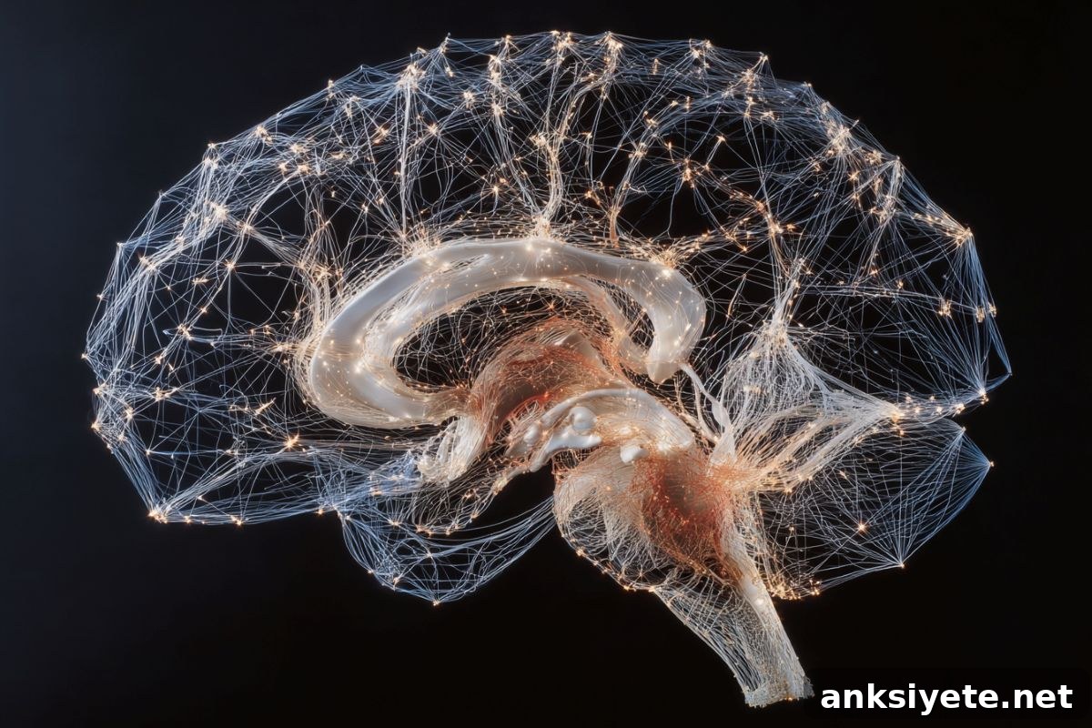
Summary: A multidisciplinary neuroengineering team has overcome a major imaging barrier by mapping the precise flow velocities of the brain’s waste-clearance network. Using physics-informed artificial intelligence to decode dynamic MRI data, the researchers revealed detailed mechanics of the glymphatic system—the fluid circulation that clears metabolic waste, including amyloid‑beta proteins associated with Alzheimer’s disease.
Their AI models identified a two-speed drainage pattern: a rapid flow along the brain’s outer surfaces and a much slower percolation through deep tissue. The outer cortical flow moves roughly 50 times faster than the deep interstitial flow.
Key Facts
- The glymphatic system: First described in 2012, this system circulates cerebrospinal and interstitial fluid around the central nervous system, especially during deep sleep, to remove metabolic byproducts that can contribute to neurodegenerative disease.
- MRI limitations: Conventional MRI provides three-dimensional anatomical images but cannot directly measure extremely slow fluid velocities occurring across the whole brain, and microscopes only capture tiny local regions.
- Physics-informed neural networks: Engineers and computational scientists created a tailored AI framework that incorporates physical laws. Trained on dynamic MRI sequences of contrast dye spreading through brain tissue, the network infers three-dimensional velocity fields, tissue permeability, and pressure estimates.
- Dual-velocity clearance: The method revealed two principal transport regimes: a faster advective flow on cortical surfaces (on the order of a few micrometers per second) and a much slower diffusion-like flow in deep brain tissue (around 0.1 μm/s), about 50 times slower.
- Clinical roadmap: Baseline flow maps so far come from animal models. The team is adapting the tools for human clinical MRI to compare fluid dynamics across ages and disease states and to develop diagnostic and monitoring applications.
- Potential impact: Mapping glymphatic circulation could enable earlier detection of impaired brain clearance tied to Alzheimer’s and provide a noninvasive test to assess fluid disruption after traumatic brain injury or concussion.
Source: University of Rochester
During deep sleep, a waterlike fluid circulates through and around the brain, flushing metabolic waste. This clearance mechanism is central to brain health and has been linked to protection against disorders such as Alzheimer’s disease.
Although researchers can visualize small patches of this circulation with microscopes, imaging whole‑brain flow in living subjects has remained elusive because the movements are extremely slow and lie below the direct sensing range of conventional MRI.
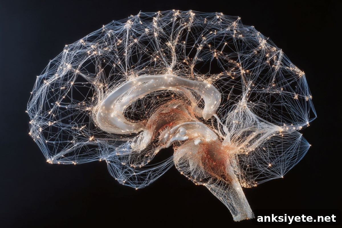
Professor Douglas Kelley (Mechanical Engineering, University of Rochester) explains the tradeoff: microscopes offer unmatched spatial detail over tiny regions, while MRI covers the whole brain but historically cannot resolve the very slow velocities involved. To bridge that gap, the research team developed MR-AIV—magnetic resonance artificial intelligence velocimetry—a physics-informed AI framework that reconstructs three-dimensional velocity fields from dynamic contrast-enhanced MRI sequences.
By feeding time-resolved dye-distribution videos into the physics-guided neural network, MR-AIV infers how rapidly fluid moves in different compartments and estimates tissue permeability and pressure fields that other methods cannot retrieve. Applied to experimental brains, this approach uncovered a functional landscape of interstitial and perivascular transport, quantitatively separating diffusion-dominated transport (~0.1 μm/s) from faster advective flow (~3 μm/s).
So far, work has focused on animal models to establish baselines and validate the method. The team is now refining the software and acquisition protocols for human clinical MRI so clinicians can compare fluid dynamics across healthy and diseased populations and across age groups.
Kelley notes several promising clinical applications: identifying impaired brain clearance in patients at risk for Alzheimer’s, screening asymptomatic individuals for poor glymphatic circulation, and assessing whether concussion or other head trauma has disrupted a patient’s internal fluid pathways to guide individualized recovery.
Funding: Supported by the NIH National Center for Complementary and Integrative Health and the NIH BRAIN Initiative.
Key collaborators on the study include Juan Diego Toscano (Brown University), Yisen Guo (University of Rochester), Zhibo Wang (Brown University), Mohammad Vaezi (University of Rochester), Yuki Mori (University of Copenhagen), George Karniadakis (Brown University), and Kimberly Boster (University of Rochester).
Key Questions Answered:
Q: Why does the brain circulate a waterlike fluid during deep sleep?
A: Deep sleep opens pathways for the glymphatic system so cerebrospinal and interstitial fluid can wash away metabolic waste produced during waking activity. Clearing toxic molecules such as amyloid‑beta reduces long‑term risk for neurodegenerative conditions.
Q: Why was artificial intelligence needed if MRIs are already in clinical use?
A: Standard MRI provides excellent structural detail but cannot directly measure the extremely slow velocities of glymphatic transport. Physics-informed AI combines MRI dynamics with physical constraints to infer flow velocities and tissue properties that the raw images cannot reveal.
Q: How could a brain fluid-flow map help after a concussion?
A: Concussive impacts can disrupt the glymphatic pathways and impair the brain’s clearance function. An AI-derived flow map would offer a rapid, noninvasive assessment of whether circulation is compromised, helping tailor recovery and return-to-play decisions.
Editorial Notes:
- This article was edited by a Neuroscience News editor.
- Journal paper reviewed in full by editorial staff.
- Additional context added by the newsroom to clarify technical details.
About this neuroscience and AI research news
Author: Luke Auburn
Source: University of Rochester
Contact: Luke Auburn – University of Rochester
Image credit: Neuroscience News
Original Research (open access): MR-AIV reveals in vivo brain-wide fluid flow with physics-informed AI — Juan Diego Toscano, Yisen Guo, Zhibo Wang, Mohammad Vaezi, Yuki Mori, George Em Karniadakis, Kimberly A. S. Boster, and Douglas H. Kelley. Science Advances. DOI: 10.1126/sciadv.aeb0404
Abstract (summary)
MR-AIV reveals in vivo brain-wide fluid flow with physics-informed AI
Cerebrospinal and interstitial fluid circulation is essential to clear metabolic waste from the brain, and disruptions to this circulation are linked to neurological disorders. Measuring brain-wide fluid transport in vivo, particularly in deep regions, has been challenging. MR-AIV is a physics-informed AI framework that reconstructs three-dimensional velocity fields, estimates tissue permeability and pressure, and reveals functional patterns of interstitial and perivascular flow. Applied to dynamic contrast-enhanced MRI, MR-AIV distinguishes slow, diffusion-dominated transport (~0.1 μm/s) from faster advective flow (~3 μm/s), enabling new investigations of brain clearance mechanisms in health and disease and offering broader applications for porous media systems.