
Summary:
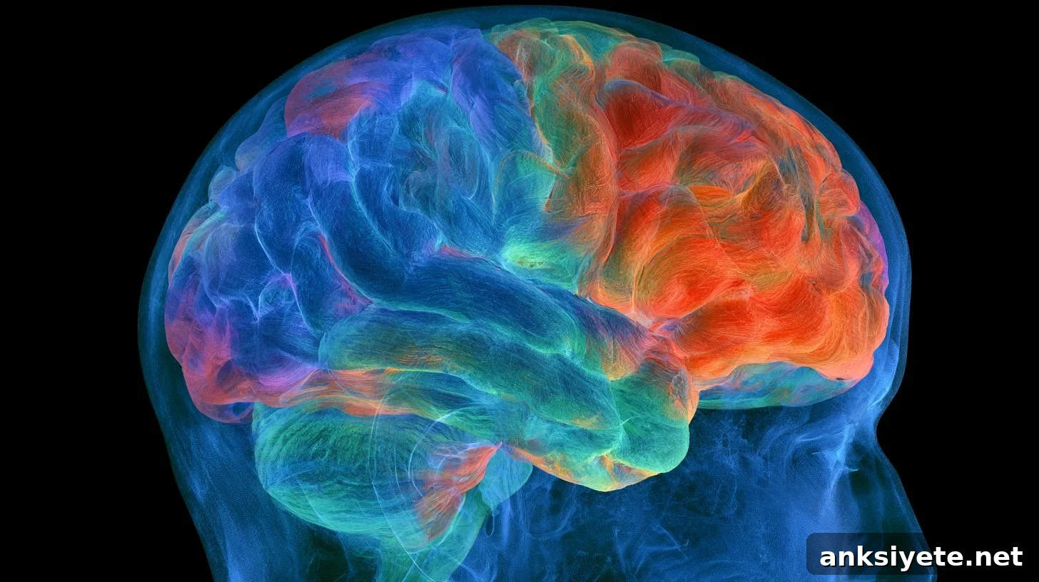
A new study finds that accelerated biological aging in brain regions spared by a stroke plays a major role in language impairment and long-term recovery. Structural measures of brain “age” in uninjured tissue predicted the severity of aphasia and forecasted language recovery six months after patients received speech therapy combined with noninvasive brain stimulation.
Key Facts:
- Impact of Intact Tissue: Biological aging patterns in the hemisphere opposite the stroke lesion explained aphasia severity independently of the lesion’s size or precise location.
- Predicting Rehabilitation Outcomes: Structural brain-age metrics taken before treatment reliably predicted language improvement measured six months after intensive speech therapy paired with noninvasive brain stimulation.
- Feasible Clinical Tool: The predictive approach uses routine, non-contrast clinical MRI scans analyzed by a free, open-access machine-learning tool trained on normative human aging data, making it accessible for many clinical and research settings.
Source: Society for Neuroscience / University of South Carolina Floyd School of Medicine
Background: After an ischemic or hemorrhagic stroke, damage is rarely confined to the immediate lesion. Areas of the brain that avoid direct ischemic injury can nonetheless show signs of accelerated structural aging. This secondary vulnerability is particularly relevant to post-stroke aphasia, a debilitating language disorder with wide variability in initial severity and in patients’ capacity to benefit from rehabilitation.
Traditionally, clinicians have tried to predict recovery by measuring focal damage—lesion volume, lesion location, and disruption of known language pathways. Yet these measures often cannot account for why two patients with very similar lesions recover so differently. The new work, published in The Journal of Neuroscience and led by Nicholas Riccardi, Leonardo Bonilha, and colleagues at the University of South Carolina Floyd School of Medicine, shows that the condition of the remaining, uninjured brain tissue is a crucial determinant of language outcomes.
Machine Learning and the “Brain Age” Gap
To detect subtle, widespread structural changes, the researchers applied an online machine-learning platform trained on large, normative datasets of human brain aging. This model compares an individual’s structural MRI to expected benchmarks for chronological age and computes a brain-age gap that reflects accelerated or decelerated structural aging relative to peers.
The study evaluated 188 post-stroke patients with varying levels of aphasia. Markers of accelerated structural aging in the hemisphere not directly affected by the stroke strongly correlated with aphasia severity, independent of classic lesion-based predictors. In other words, the biological condition of the uninjured hemisphere provided additional explanatory power for language impairment beyond what lesion maps alone could offer.
The investigators also followed a subgroup of patients who completed an intensive rehabilitation protocol combining speech-language therapy with noninvasive brain stimulation. Baseline brain-age measures taken before therapy successfully predicted the magnitude of sustained language gains assessed six months after the intervention, suggesting these metrics have prognostic value for long-term recovery.
Accessible, Low-Cost Biomarkers for Rehabilitation
A major strength of this approach is its accessibility. The predictive framework relies on routine structural MRI and a free, open-access computational algorithm, avoiding expensive or technically demanding imaging sequences. That lowers barriers to implementation and could enable clinicians and researchers in diverse settings to generate objective biomarkers that support personalized rehabilitation planning.
Lead author Nicholas Riccardi emphasized the practical implications: “This work suggests that recovery potential after stroke depends on the health of the rest of the brain, which is partly shaped by treatable factors like cardiovascular health. Second, because everything here came from a single routine scan and a free online tool, this could realistically reach a variety of clinical or research settings one day.”
The findings point to modifiable systemic factors—blood pressure, metabolic health, physical activity and similar influences—that could help preserve or improve global brain resilience. Protecting the health of uninjured neural networks may make them more capable of supporting neuroplasticity and functional recovery after stroke.
Editorial Notes:
- This article was edited by a Neuroscience News editor.
- The full journal paper will be reviewed upon its release.
- Additional context was provided by our editorial staff.
About this Neurology Research:
- Media Contact: SfN Media
- Source: SfN
- Image Credit: Image credited to Neuroscience News
- Original Research: The findings will be published in The Journal of Neuroscience (open access).