
Summary:
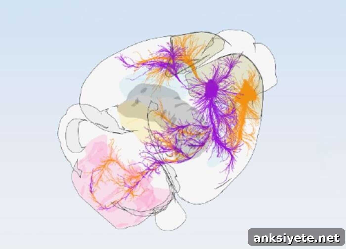
An international team of researchers has produced the most detailed map to date of the mouse motor cortex, identifying 16 distinct subregions arranged along two anatomical axes rather than the traditional two-zone model. Their findings show that primary and secondary motor areas operate in parallel, not in a strict hierarchy, and provide an open, standardized framework to study selective neuronal vulnerability in disorders such as ALS and frontotemporal dementia.
Key Facts:
- Sixteen distinct subregions across two axes: Analysis of 547 projection-tracing experiments revealed 16 modules organized into three rows, with an anterior–posterior axis separating planning-related regions from sensory-integrative regions, and a medial–lateral axis reflecting somatotopic representations from trunk and limbs to face and mouth.
- Parallel organization, not hierarchy: Primary and secondary motor cortices send projections in parallel to brainstem and spinal cord targets, contradicting the long-held model that secondary motor areas mainly relay signals through primary motor cortex.
- Multimodal validation and open access: The 16-module blueprint was validated using single-neuron reconstructions, projection patterns, and cell-type mapping. The atlas has been integrated into the BrainGlobe framework for broad research use.
Source: University of Basel / Allen Institute / Friedrich Miescher Institute for Biomedical Research
The motor cortex sits at the center of voluntary movement, directing everything from precise finger movements to chewing and locomotion. For decades standard atlases simplified this region into two broad compartments: the primary motor cortex (M1) and the secondary motor cortex (M2). That coarse division failed to capture the fine-grained specializations, diverse cell types, and precise connection patterns that underlie motor behavior.
To refine that picture, researchers from the University of Basel, the Friedrich Miescher Institute for Biomedical Research, and the Allen Institute systematically mapped how cortical outputs distribute across the brain. By clustering cortical locations with shared projection targets, they defined 16 projection-defined modules, each with a characteristic wiring signature.
“A precise wiring map is essential for understanding disease and designing treatments,” said Hongkui Zeng of the Allen Institute, senior author of the study. Combining detailed tracing data with expert anatomical mapping produced a high-resolution blueprint of the circuits that control movement.
A Dual-Axis Coordinate System: 16 Motor Modules
The team analyzed 547 projection-tracing experiments from the Allen Institute’s Mouse Brain Atlas to chart where small cortical regions send outputs across sensory, motor, and cognitive targets. Clustering areas by shared targets revealed 16 discrete modules arranged along two principal biological axes:
- Anterior–Posterior Axis: Anterior modules are enriched for higher-level motor planning and decision-making connections, while posterior modules align closely with sensory feedback pathways such as touch and proprioception.
- Medial–Lateral Axis: This axis follows somatotopy, transitioning from trunk and limb representations medially to face, jaw, and mouth regions laterally.
The 16-region architecture was validated using independent approaches: detailed reconstructions of individual projecting neurons and spatial profiling of diverse cortical cell types. All methods converged on the same modular layout, supporting a robust and biologically meaningful parcellation.
Importantly, the study resolves a long-standing question about cortical hierarchy. Rather than acting in series, M1 and M2 send parallel projections to brainstem and spinal cord targets, implying coordinated, concurrent routes for motor commands instead of a strict top-down relay.
“We were struck by the precision with which these modules target output regions and by the consistent wiring logic that governs cortical and subcortical connections,” said Silvia Arber, lead author and professor of neurobiology.
Implications for ALS, FTD, and Disease Mapping
The open-access 16-subregion blueprint offers immediate utility for studying neurodegenerative diseases that affect motor circuits. Amyotrophic lateral sclerosis (ALS) preferentially destroys upper and lower motor neurons, while frontotemporal dementia (FTD) primarily impacts frontal cortical networks governing behavior and language. Both disorders show selective vulnerability of particular cell populations, and the reasons for that selective loss remain unclear.
With a standardized, high-resolution map, researchers can pinpoint which anatomical compartments contain vulnerable cell types, follow how pathology spreads through defined output pathways, and compare findings across labs using a shared coordinate system. To facilitate widespread adoption, the framework has been incorporated into BrainGlobe, enabling alignment of experimental data to this modular atlas.
“Combining large-scale wiring datasets with cellular profiling revealed a more precise organizational blueprint for the motor cortex,” said co-first author Harsh Kanodia. Co-first author Antonio Falasconi added that the unified map will help researchers align and interpret cortical data more effectively, accelerating progress in motor system and disease research.
Editorial Notes:
- This article was edited by a Neuroscience News editor.
- The journal paper was reviewed in full by the editorial team.
- Additional context was provided by staff writers.
About this Brain Mapping and Neuroscience Research:
- Media Contact: Peter Kim
- Source: Allen Institute
- Image Credit: Friedrich Miescher Institute for Biomedical Research, University of Basel
- Original Research (Open Access): Cell (September 23, 2026). Title: “Projection-defined modules reveal mouse motor cortex architecture.” Authors: Antonio Falasconi, Harsh Kanodia, Nicholas Lusk, Shenqin Yao, Rui M. Costa, Hongkui Zeng, and Silvia Arber.
- DOI: 10.1016/j.cell.2026.08.046
Abstract
Projection-defined modules reveal mouse motor cortex architecture
The motor cortex coordinates movement through complex connectivity, yet organizing principles of this region have remained disputed. This study demonstrates that subcortical projections from mouse motor cortex define 16 distinct modules. Differences in subcortical output align with variations in corticocortical connectivity and cell-type composition, delineating two spatial axes within the motor cortex.
Along one axis, primary motor cortex is reciprocally coupled with somatosensory areas while secondary motor cortex preferentially connects with frontal regions; distinct excitatory neuron populations help differentiate these areas. Along the orthogonal axis, somatosensory inputs and non-sensorimotor cortical wiring stratify the modules, which are further characterized by aligned cell-type signatures.
This two-axis cortical logic extends to subcortical targets: striatum, thalamus, and brainstem follow specific convergence–divergence patterns that integrate cortical inputs differently. Together, these findings reveal how the anatomical architecture of mouse motor cortex organizes into brainwide and specialized neuronal networks.