
Summary:
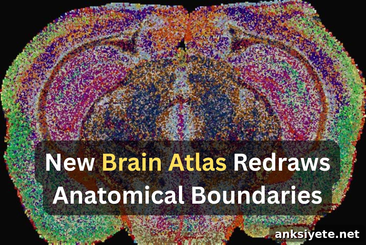
Researchers have produced the first comprehensive three-dimensional Lipid Brain Atlas of the mouse brain, mapping 539 distinct “lipizones”—chemical territories defined entirely by their lipid profiles. This high-resolution atlas shows that lipids act like molecular postal codes, linking distant but functionally related regions, and reveals unexpected chemical diversity within white matter that traditional gene-based maps have missed.
Key Facts:
- 539 Lipizones Defined: Using mass spectrometry imaging and custom machine-learning tools to process roughly seven million measurements across whole brains, the team delineated 539 metabolic territories based solely on their lipid combinations.
- Lipids as Molecular Postal Codes: Lipid distributions are highly organized and align with anatomical boundaries; they can link a neuron’s cell body to the distant terminal regions reached by its axons, providing a shared chemical identity across connected sites.
- White Matter Is Chemically Heterogeneous: What appeared to be uniform myelin-rich white matter is actually a mosaic of distinct biochemical zones, indicating that myelin-producing cells display biochemical diversity not apparent from gene expression alone.
Source: EPFL (École Polytechnique Fédérale de Lausanne)
The brain is commonly described in terms of cells and electrical connections, but a large portion of its structure is built from lipids. These fatty molecules form cell membranes, insulate axons with myelin, and influence how neurons transmit and receive signals. Despite their importance, lipids have remained difficult to map across whole tissues at high resolution, leaving a gap in our understanding of brain chemistry.
“Until now we only had a blurry view of how lipids vary across brain regions,” says Giovanni D’Angelo, professor at EPFL. “Filling that gap matters because changes in brain lipids are linked to conditions ranging from mood disorders to Alzheimer’s disease.”
A Distinct Chemical Map
An interdisciplinary team led by Giovanni D’Angelo, Luca Fusar Bassini, and Gioele La Manno at EPFL built the first detailed, high-resolution lipid atlas of the whole mouse brain. Published in Nature, the Lipid Brain Atlas demonstrates that lipid distributions form a precise biochemical architecture that corresponds closely with known anatomy.
The lipid signatures were so distinctive that researchers could identify the anatomical origin of a tissue sample from its lipid profile alone. In many cases the lipizones matched established cell-type territories and classical brain regions; in others they revealed new relationships that traditional maps had overlooked.
Building the Atlas: Mass Spectrometry Meets Machine Learning
To create the atlas, the team used mass spectrometry imaging, scanning thin brain sections point by point with a laser to record the precise lipid composition at each location. They measured 172 lipid species across 109 slices from 11 mice and combined approximately seven million data points into a coherent three-dimensional model.
Because of the dataset’s scale and complexity, the researchers developed new machine-learning tools tailored to assemble and interpret the measurements. The final map divides the brain into 539 lipizones, each defined by a unique biochemical mixture.
Beyond matching known structures, lipizones often connect cell bodies with the remote terminal fields their axons innervate. This chemical continuity across connected regions highlights organizational features that gene-based atlases, which typically concentrate on cell bodies, can miss.
“Lipids act like postal codes, assigning the same chemical address to distant but related brain sites,” D’Angelo explains.
White Matter’s Patchwork Chemistry and Pregnancy-Related Changes
One striking discovery is that white matter is not chemically uniform. Instead, it forms a patchwork of lipizones, revealing an unappreciated axis of oligodendrocyte or myelin-related heterogeneity that gene-expression maps did not capture.
The atlas also enabled the team to track lipid remodeling during pregnancy. In pregnant mice, lipid composition shifted substantially across many brain regions—changes that were larger overall than typical male–female differences. The cortex, especially outer layers, showed pronounced remodeling, while galactosyl ceramide, an important myelin component, increased across much of the white matter.
“These changes indicate the brain retunes its lipidome to meet the physiological demands of pregnancy,” says Gioele La Manno. “The atlas shows how lipids adapt to changing bodily needs.”
A Public Resource to Advance Disease Research
The Lipid Brain Atlas is available as an open resource for the research community, providing a healthy reference map of brain lipid organization. Researchers can use this baseline to localize where lipid metabolism deviates in neurodevelopmental disorders, aging, or neurodegeneration.
“What we can transfer to human studies is the approach and the principle that a healthy brain has an organized lipid map,” Luca Fusar Bassini notes.
Editorial Notes:
- This article was edited by a Neuroscience News editor.
- The journal paper was reviewed in full.
- Additional context was provided by staff.
About this Genetics and Neuroregeneration Research:
- Media Contact: Gioele La Manno
- Source: EPFL
- Image Credit: Image credited to EPFL/Luca Fusar Bassini
- Original Research is Open Access: Nature (September 23, 2026). “The lipidomic architecture of the mouse brain.” Authors: Luca Fusar Bassini, Halima Hannah Schede, Laura Capolupo, Leila Haj Abdullah Alieh, Irmak Kaysudu, Francesca Venturi, Hannah Hochgerner, Alessandro Valente, Colas Droin, Daniel Trejo Banos, Irina Khven, Jean Andrea Maillat, Anne-Laure Mahul-Mellier, Antonino Asaro, Doğukan H. Ülgen, Pavel Barahtjan, Ece Z. Asirim, Anita Nasrallah, Carmen Sandi, Ekaterina Krymova, Giovanni D’Angelo & Gioele La Manno.
- DOI: 10.1038/s41586-026-11050-0
Abstract
The lipidomic architecture of the mouse brain
Lipids are essential to brain structure and function, supporting synaptic transmission and signal propagation. Changes in lipid composition are implicated in many neurological conditions, yet the spatial organization of the brain lipidome has been less well characterized than other molecular modalities.
Here, we mapped membrane lipids across the adult mouse brain at micrometric resolution, comparing sexes and examining pregnancy. The resulting lipid brain atlas shows a fine-grained biochemical structure that aligns with functional anatomy.
Spatial heterogeneity in membrane lipids clusters into territories called “lipizones.” Lipizones partially reflect known cell-type distributions but also capture distal axon terminals. Using lipizones, we (1) reveal principles of grey matter lipid organization related to connectivity and cytoarchitecture; (2) identify a new axis of oligodendrocyte-related heterogeneity within white matter; and (3) detect biochemical zonation in the choroid plexus and ventricular walls.
We show that this lipidomic architecture adapts to physiological change: in pregnant female mice the white matter becomes metabolically active and cortical lipizones undergo region-specific remodeling, notably in layer 4.
These results provide a foundational resource (https://lbae-v2.epfl.ch/) that will help reshape understanding of lipids in brain development, physiology, and disease.