
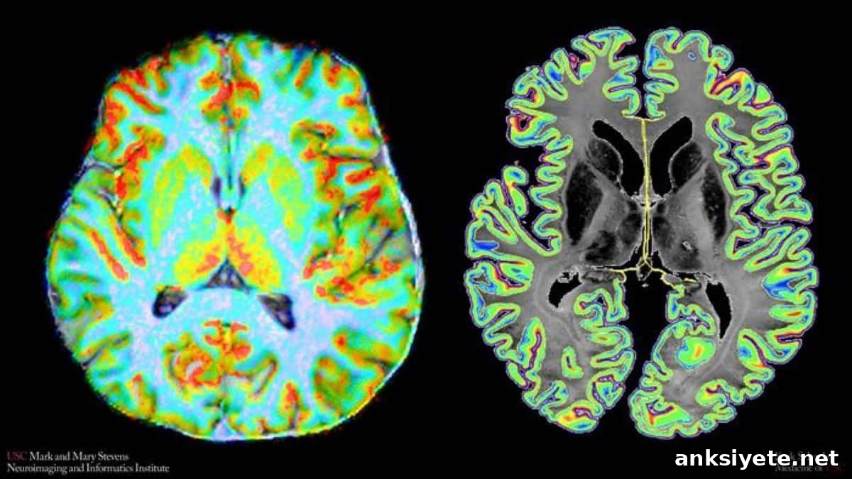
Summary:
Researchers at the Keck School of Medicine of USC have developed a noninvasive metric, the cerebral blood flow–cell-body staining intensity similarity index (CCSI), to quantify how laminar blood flow aligns with cellular density across the living human cerebral cortex. Combining ultra-high-field 7-Tesla MRI with detailed cellular and molecular atlases, the team shows that tight vascular–cellular alignment corresponds to higher mitochondrial respiratory capacity and improves predictions of higher-order cognitive function.
Key Facts:
- Novel laminar metric: CCSI uses 7-Tesla arterial spin labeling (ASL) MRI together with the 3D BigBrain cell-body staining atlas to measure how closely laminar perfusion mirrors cellular packing across 360 cortical parcels.
- Metabolic capacity, not volume: Stronger alignment between blood flow and cell density is linked to greater mitochondrial respiratory capacity per mitochondrion, rather than simply larger mitochondrial volume or bulk blood flow.
- Improved prediction of cognition: Adding CCSI to structure–function coupling models significantly enhances prediction of neural activity in higher-order association areas involved in memory, attention, and reasoning.
Source: Keck School of Medicine of USC
The cerebral cortex is organized into distinct cellular layers, each with varying densities and types of neurons and glia. Neural tissue has almost no intrinsic energy reserve, so maintaining function depends on a continuous, tightly regulated supply of oxygen and glucose delivered by cerebral blood vessels.
Traditional neuroimaging approaches typically average vascular signals across the full cortical thickness, masking how microvascular perfusion supports the metabolic needs of specific laminar compartments. To overcome this limitation, investigators at the Mark and Mary Stevens Neuroimaging and Informatics Institute (Stevens INI) developed CCSI, a reproducible, noninvasive index that links laminar perfusion profiles to cortical cytoarchitecture.
Their findings, published in Nature Communications, provide a tool for visualizing and quantifying how microvascular perfusion is organized relative to cellular architecture in vivo.
“Because the brain can’t store much energy, its cells depend on a precise and continuous blood supply,” said Fanhua Guo, co–first author and researcher at Stevens INI. “CCSI gives us a way to assess whether that supply is positioned to meet layer-specific cellular demands across the cortex.”
Ultra-High-Field Laminar Perfusion
To resolve perfusion across cortical layers in living participants, the team used arterial spin labeling (ASL) on a 7 Tesla MRI scanner. ASL magnetically labels arterial water, using it as an endogenous tracer to measure perfusion at an isotropic 1 mm resolution. The study scanned 30 healthy adults, with 14 returning for repeat sessions to verify test–retest reliability.
Researchers parceled the cortex into 360 regions and tracked perfusion from the outer cortical surface to deeper layers, comparing laminar blood flow profiles with cell-body staining intensity derived from BigBrain, a high-resolution 3D histological reconstruction of human cortical cytoarchitecture.
Results showed that blood flow and cellular density were aligned across most cortical regions: layers with greater cell packing generally received proportionally more perfusion. This vascular–cellular coupling was strongest in primary sensorimotor and visual cortices, areas specialized for basic sensory processing and motor control.
Connecting Perfusion to Mitochondrial Power and Glia
To investigate biological determinants of CCSI, the authors integrated laminar perfusion measures with independent maps of mitochondrial function, single-cell transcriptomics, and regional gene expression. CCSI correlated selectively with mitochondrial respiratory capacity — the maximal ATP production rate per mitochondrion — rather than total mitochondrial volume or conventional bulk blood flow.
At the cellular level, CCSI aligned closely with capillary endothelial populations, which regulate local microperfusion and form the blood–brain barrier, and with mature oligodendrocytes, which both insulate axons and provide metabolic support to neuronal processes. Transcriptomic analyses linked high-CCSI regions to gene networks involved in angiogenesis, energy metabolism, and mitochondrial maintenance.
Decoding Higher-Order Cognitive Circuits
The investigators also tested whether CCSI could improve structure–function coupling, a persistent challenge in systems neuroscience. While structural anatomy reliably predicts function in primary sensory and motor areas, higher-order association cortices—responsible for attention, memory, and complex reasoning—often show weaker correspondence between structure and function.
When CCSI was incorporated into structure–function models, prediction of functional activity in association networks improved markedly. This suggests that regional organization of vasculature and metabolic capacity contributes to neural computations in ways that morphology alone does not capture.
Although current CCSI analyses focus on group-level patterns and rely on postmortem atlases for molecular interpretation, future work aims to apply CCSI to individual patients. Disruptions of cerebral microcirculation, mitochondrial energy production, and oligodendrocyte function are implicated in conditions such as Alzheimer’s disease, multiple sclerosis, and schizophrenia. The authors propose that CCSI could ultimately serve as a biomarker to detect early neurovascular and metabolic dysfunction and to evaluate therapeutic strategies.
About the study
The study team includes Fanhua Guo, Chenyang Zhao, Danny JJ Wang, Ravi R. Bhatt, Zixuan Liu, Zidong Yang, Kay Jann, Xingfeng Shao, Neda Jahanshad, Mara Mather, Andy Jeesu Kim, and Siyi Xu, among others from Stevens INI, USC Leonard Davis School of Gerontology, USC Department of Psychology, USC Department of Biomedical Engineering, and the University of Washington.
Funding: Supported by the National Institutes of Health under grants UF1-NS100614, S10-OD025312, R01-EB032169, RF1-AG084072, R01-MH134004 and R01-NS134712.
Editorial Notes:
- This article was edited by a Neuroscience News editor.
- The journal paper was reviewed in full.
- Additional context was added by staff.
About this Genetics and Neurology Research:
- Media Contact: Laura LeBlanc
- Source: USC
- Image Credit: Stevens INI
- Original Research (Open Access): Nature Communications (September 15, 2026). Title: “Assessing molecular, cellular and transcriptomic bases of laminar perfusion and cytoarchitecture coupling in the human cortex.” Authors: Fanhua Guo, Chenyang Zhao, Ravi R. Bhatt, Zixuan Liu, Andy Jeesu Kim, Zidong Yang, Siyi Xu, Kay Jann, Xingfeng Shao, Mara Mather, Neda Jahanshad & Danny JJ Wang.
- DOI: 10.1038/s41467-026-76812-w
Abstract
Assessing molecular, cellular and transcriptomic bases of laminar perfusion and cytoarchitecture coupling in the human cortex
Understanding how cellular architecture shapes cortical function requires mesoscopic approaches that resolve structure–function relationships in vivo. We introduce the cerebral blood flow (CBF)–cell-body staining intensity (CSI) similarity index (CCSI), a quantitative measure of laminar perfusion–cytoarchitecture coupling based on whole-brain 1 mm isotropic 7 T arterial spin labeling and BigBrain cell-body staining profiles.
Across 30 participants, CCSI revealed reproducible, region-specific alignment between laminar perfusion and cellular density. CCSI associated selectively with mitochondrial respiratory capacity per mitochondrion and colocalized with capillary endothelial and mature oligodendrocyte populations. Gene ontology enrichment implicated metabolic regulation, neurovascular organization, and mitochondrial homeostasis. A control analysis using quantitative T1 showed no significant associations, supporting the specificity of the findings. At the systems level, CCSI improved structure–function gradient correspondence in higher-hierarchy association cortices.
These results establish CCSI as a reproducible, noninvasive mesoscopic index linking cortical perfusion, microstructure, and metabolism.