Summary: Researchers report that maternal immune activation during pregnancy reshapes the epigenome of developing fetal brain cells, interfering with the formation of deep-layer cortical neurons and raising the likelihood of neurodevelopmental disorders such as autism.
Key Facts:
- Epigenetic Alterations: Maternal immune activation (MIA) changes DNA methylation patterns in the fetal frontal cortex, producing hypermethylation at genomic sites where the transcription factor Tbr1 normally binds—effectively blocking Tbr1 from executing its role in deep-layer neuron development.
- Autism Overlap: Roughly 25% of high-confidence autism-linked genes in the SFARI Gene Database were dysregulated in deep-layer neurons from offspring exposed to MIA in this study.
- Lasting Brain Circuitry Changes: The prenatal immune challenge reshaped fetal gene expression and methylation, causing impaired neurodevelopment and electrophysiological abnormalities that persisted into adulthood.
Source: Salk Institute
Neurodevelopmental conditions such as autism spectrum disorder (ASD) and attention-deficit/hyperactivity disorder (ADHD) affect a significant portion of the population. While the precise origins of many of these conditions remain complex and multifactorial, epidemiological data have long linked severe maternal infection during pregnancy with an increased risk of neurodevelopmental disorders in children.
Scientists at the Salk Institute have now mapped a cellular and molecular pathway that helps explain this association. Using a well-established mouse model of maternal immune activation—where pregnant mice are exposed to the viral mimetic Poly(I:C) to simulate influenza-like immune responses—the researchers examined the epigenomic and transcriptomic trajectories of frontal cortex neurons from mid-gestation through early postnatal development.
Their analysis revealed thousands of sites across the genome where DNA methylation and gene expression diverged between offspring of healthy pregnancies and those affected by MIA. These epigenetic changes were not uniformly distributed but were concentrated in regions that control the development of deep-layer excitatory neurons in the cerebral cortex.
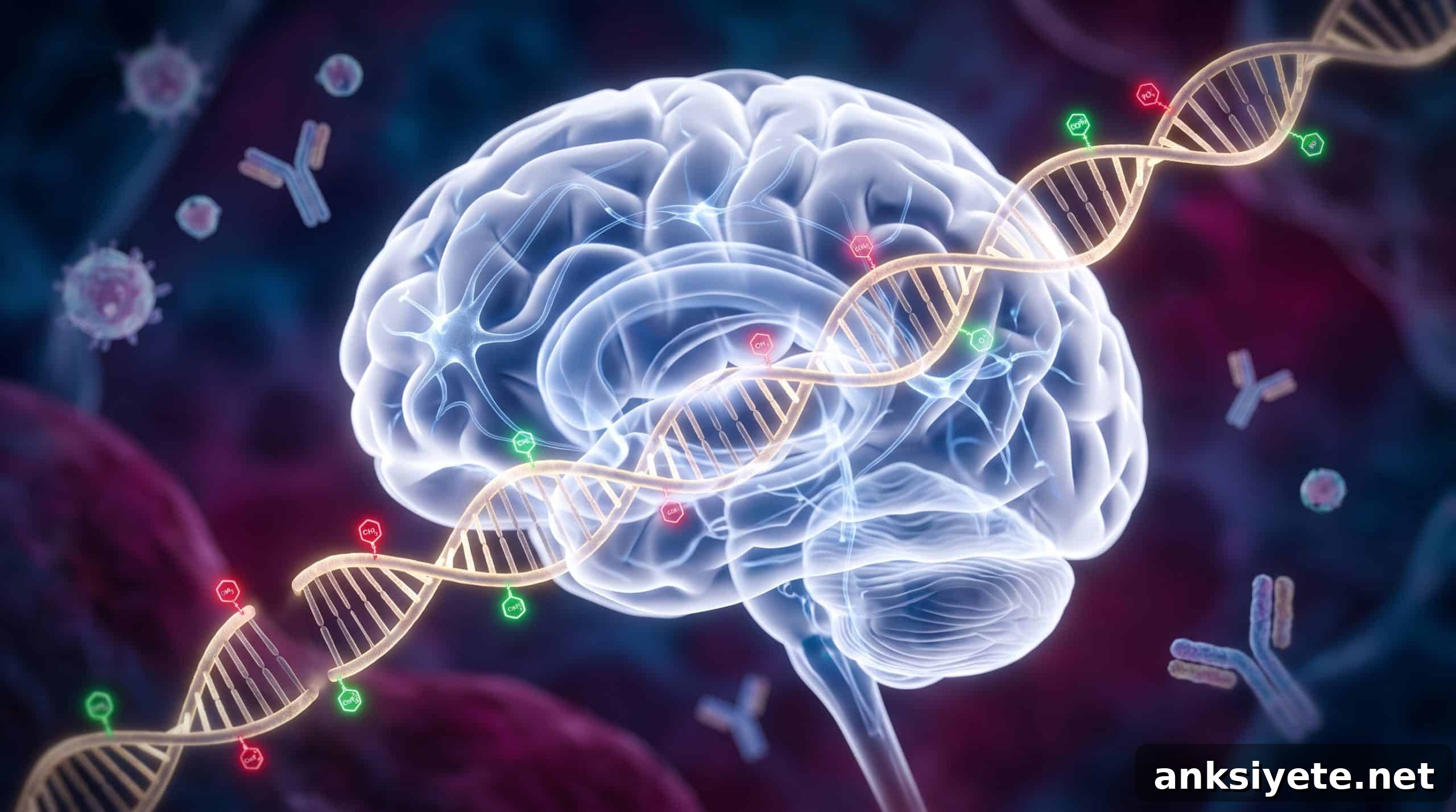
A striking finding was the hypermethylation of genomic regions that normally bind Tbr1, a transcription factor essential for specifying deep-layer neuronal identity. Although Tbr1 RNA and protein levels were present—indeed Tbr1 transcription was upregulated in some cases—the excessive methylation at its target sites prevented effective binding and downstream activation of Tbr1-regulated genes. As a result, many Tbr1 target genes were downregulated at birth, indicating that expression of the regulator itself became uncoupled from its functional influence on gene networks that shape deep-layer neurons.
The study connected these molecular signatures to functional outcomes. Electrophysiological recordings from affected offspring showed persistent alterations in the intrinsic and firing properties of deep-layer neurons, consistent with disrupted circuit maturation. These physiological changes, detected beyond the neonatal period, suggest that prenatal immune challenges can produce long-lasting modifications in cortical circuitry.
Jessica Arzavala, a co-first author and graduate researcher at Salk, noted that cross-referencing their data with the SFARI Gene Database highlighted potential relevance to autism: about one-quarter of the high-confidence autism-associated genes overlapped with dysregulated genes in deep-layer neurons from MIA-exposed offspring.
Co-corresponding author Dr. Joseph Ecker emphasized an important nuance: maternal infection alters risk but does not determine outcome in every case. “Infection changes the odds of whether neurodevelopment is affected—not everyone who gets sick during pregnancy is going to definitively have a child with a neurodevelopmental disorder,” he said, underlining that MIA is one of multiple factors that can influence neurodevelopmental trajectories.
By precisely locating the epigenetic “roadblocks” that interfere with transcription factor function during critical windows of brain maturation, the research opens pathways for future work aimed at therapeutic intervention. Mapping when and where methylation changes appear creates opportunities to explore maternal or fetal-targeted treatments designed to prevent or reduce the downstream impact on cortical development.
Co-corresponding author Dr. Margarita Behrens added that these results represent an initial but meaningful advance. “It’s just the tip of the iceberg,” she said, noting that the refined epigenomic maps will let researchers tackle more detailed questions about timing, cell-type specificity, and potential interventions.
Editorial Notes:
- This article was edited by a Neuroscience News editor.
- Journal paper reviewed in full by the editorial team.
- Additional context added by staff to clarify implications.
About this Neurodevelopment Research:
- Media Contact: Isabella Davis
- Source: Salk Institute
- Image Credit: Image generated for Neuroscience News
- Original Research is Open Access: Molecular Psychiatry (September 2, 2026). “Poly(I:C) maternal immune activation alters epigenetic programming in the developing frontal cortex.” Authors: Chi-Yu Lai, Jessica Arzavala, Antonio Pinto-Duarte, Hanqing Liu, Julia Osteen, Rosa Gomez Castanon, Joseph Nery, Shiyuan Wang, Junhao Li, Susan B. Powell, Eran Mukamel, Margarita Behrens, and Joseph Ecker.
- DOI: 10.1038/s41380-026-03856-1
Abstract
Lai, CY., Arzavala, J., Pinto-Duarte, A. et al. Maternal immune activation disrupts epigenomic and functional maturation of cortical excitatory neurons. Mol Psychiatry (2026).
Elevated maternal pro-inflammatory cytokines following severe infection during gestation can disrupt neural development in offspring and increase risk for neurodevelopmental disorders. The viral mimetic Poly(I:C) reproduces many effects of gestational influenza exposure, producing behavioral outcomes that model aspects of neurodevelopmental disorders. Although Poly(I:C)-induced maternal immune activation (PIC-MIA) is known to alter the epigenome, behavior, and cognition of offspring, the timing and cellular specificity of these changes have been unclear. In this study, investigators examined PIC-MIA effects on epigenomic maturation of the frontal cortex, focusing on excitatory neuron-specific DNA methylation and transcriptomic dynamics across perinatal development. Mid-gestation PIC-MIA disrupted the excitatory neuron transcriptome with the largest changes observed at birth. The mature DNA methylation program of excitatory neurons was altered at thousands of genomic regulatory regions that normally gain or lose methylation during development. Transcription factor binding site analysis showed significant enrichment of Tbr1 motifs within hypermethylated, deep-layer neuron-specific regions at birth. Notably, Tbr1 transcriptional targets were downregulated at birth despite increased Tbr1 transcription, suggesting that PIC-MIA uncouples Tbr1 expression from its regulatory function in deep-layer neurons. Electrophysiological recordings further confirmed lasting disruption of deep-layer neuronal activity. These results suggest that mid-gestation MIA may alter deep-layer neuron development through an epigenomic blockade of Tbr1 function, perturbing normal cortical circuit formation.